Type 1 diabetes mellitus (T1D) results from the autoimmune damage to β-cells in the pancreatic islets. Destruction of β-cells leads to insulin deficiency and, consequently, to hyperglycemia and other severe metabolic disorders. All T1D patients need lifelong insulin therapy. T1D affects mostly children and adolescents. There is a hereditary risk of T1D: close relatives of patients have approximately 40 times higher risk of the disease [1].
T1D is characterized by latent preclinical stage (PS), in which the gradual β-cell destruction takes place [2]. PS lasts months to years and ends when the population of β-cells is reduced by 70–80%. At this moment absolute insulin deficiency, hyperglycemia, and its symptoms emerge: the T1D onset occurs and the clinical stage of the disease begins. Approximately 50% of patients develop an acute T1D complication at the onset: diabetic ketoacidosis leading to severe neurocognitive disorders and eventually to death. The delayed insulin prescription is the main cause of ketoacidosis.
The idea that T1D is an autoimmune disease had erased by early 1980s. By that time it had been shown that the majority of T1D patients carry serum autoantibodies (autoAB) binding islet cell structures on the cryostat sections of the pancreas [3]. Such autoAb were named islet cell antibodies (ICA). It was clear that ICA bind to some cytoplasmic antigens of β-cells, the potential autoimmune response targets. The most likely candidate for such antigens seemed to be insulin, the main product of β-cells. This hypothesis was confirmed in 1983 by the group of the US researchers led by Jerry Palmer [4]. Palmer and his colleagues found IAA in patients with the new-onset T1D never treated by insulin, and in some healthy relatives of T1D patients by RIA.
Later, autoABs against other β-cell antigens were discovered, specifically the glutamic acid decarboxylase antibodies (GADA), islet antigen-2 antibodies (IA-2A), and zinc transporter 8 antibodies (ZnT8A) [5]. AutoABs do not play any essential role in destruction of β-cells, but serve its highly specific laboratory markers.
Testing for autoABs is used for:
- early diagnosis of T1D in the PS;
- confirmation of the T1D diagnosis when the clinical picture is confusing;
- differential diagnosis between T1D and other DM types and variants;
- screening for the T1D PS in persons at risk (for example, among the first-degree relatives) and in the population.
The latter task is of special importance for two reasons. First, the detection of the β-cell destruction markers suggests high probability of T1D onset and allows the patients and their parents to get ready. It also allows physicians to timely prescribe insulin therapy and prevent ketoacidosis and its sequelae. Second, screening reveals the patients having indications for drug prevention of T1D involving the use of the drug suppressing the anti-β-cell immune response, for example teplizumab [6].
The screening programs have been conducted for many years in European countries, the USA, Canada, Australia, and Israel [7], and in the end of 2024 such a program was launched in Russia, in the Endocrinology Research Center [8]. The tests for autoABs represent the main screening tool, and the central place is occupied by the test for IAA, since this autoAB emerges as early as in the beginning of the PS and serves as the earliest indicator of the anti-β-cell immune response [9].
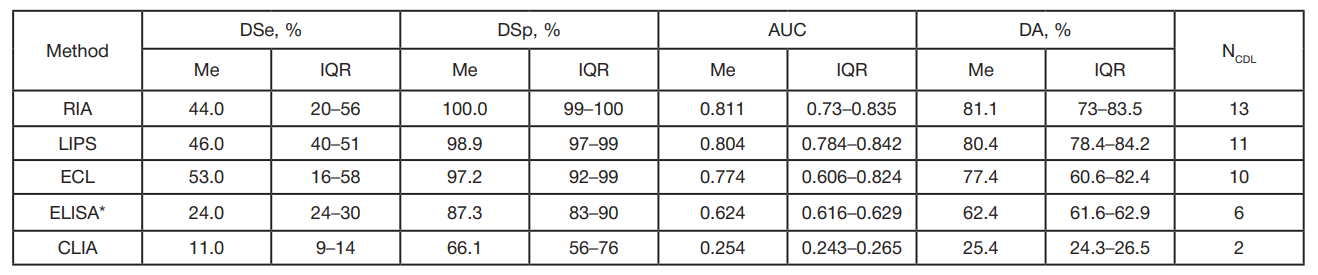
Various methods are used for IAA testing in different labs. The most common ones are RIA, LIPS (Luciferase Immunoprecipitation System) assay; electrochemiluminescence (ECL) analysis; ELISA and CLIA (chemiluminescent immunoassay). Performance characteristics of these methods, diagnostic sensitivity (DSe), diagnostic specificity (DSp), and diagnostic accuracy (DA), differ considerably. Comparative assessment of different IAA tests is periodically conducted as part of the international Islet Autoantibody Standardization Program (IASP) [10]. The participating labs receive the sets of sera from patients with new-onset T1D and from healthy blood donors; each lab performs testing of all sera for IAA by its own method. The results of two IASP rounds conducted in 2018 and 2020 are provided in tab. 1.
As can be seen, RIA yields the best DSe and DSp, along with the maximum DA; LIPS assay ranks second in DA, ELISA ranks fourth, and CLIA shows no DA (AUC < 0.5). The ELISA and CLIA unsatisfactory characteristics are explained by the fact that in these methods an antigen (insulin) is absorbed on the solid phase (plastic or magnetic particles), which leads to disruption of its conformation and shielding of antigenic determinants.
Unfortunately, absolutely all Russian labs use the commercially available ELISA systems for IAA testing. The operating parameters of those are even worse than that of ELISA systems represented in the IASP. For example, in the widely used Orgentec Anti-Insulin kit (Orgentec Diagnostika GmbH, REF ORG520, Germany) DSe = 4%, DSp = 95.6%, DA = 50%, i.e. its results are of no clinical significance [11].
Recently, the Maglumi IAA CLIA system (Shenzhen New Industries Biomedical Engineering Co., Ltd; China) became available on the Russian market, but the system user manual contains no data on its performance characteristics [12].
Thus, in our country there is a need to develop and introduce reliable, informative test system for IAA determination. In this regard, the Russian Children's Clinical Hospital diagnostic lab attempted to reproduce classical RIA IAA.
METHODS
Study overview
Serum samples from the patients with the maximum and minimum likelihood of being IAA carriers, i.e. patients with new-onset T1D (T1D group) and patients having no such disorder (group C, control), were tested for IAA. The study was conducted in January—February 2024 in the Russian Children's Clinical Hospital and Institute of Bioorganic Chemistry.
Description of patient groups
T1D group (n = 21)
M : F = 8 : 13 (38% : 62%); age 1.1–17.9 years (median age 10.1 years, 95% confidence interval for the median 4.2–12.1 years).
Inclusion criteria: age 0–18 years; diagnosis “type 1 diabetes mellitus, new-onset” (ICD-10 E10.1 or E10.9); T1D duration from the date of the diagnosis to the date of blood sample collection ≤ 3 months; presence of at least two autoAB types out of the following: ICA, GADA, IA-2A, ZnT8A.
Group C (n = 19)
M : F = 12 : 7 (63% : 37%); age 2.5–46.9 years (median age 13.3 years, 95% confidence interval for the median 10.2–16.6 years).
Inclusion criteria for the group: any age, any sex; the patient is generally healthy (ICD-10 Z00) or diagnosed with one of the follwing: type 2 diabetes mellitus (ICD-10 E11), other specified diabetes mellitus forms, including various monogenic DM forms (ICD-10 E13, E13.9), obesity (ICD-10 E66), unspecified DM (ICD-10 E14, E14.9), Cushing syndrome (ICD-10 E24), pituitary-dependent Cushing's disease (ICD-10 E24.0), Turner syndrome (ICD-10 Q96); patient was never diagnosed with T1D; patient never received insulin injections; no GADA, IA-2A, ZnT8A in the patient’s serum.
IAA testing method
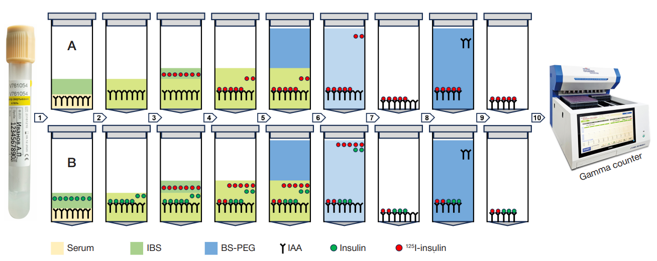
Competitive radioimmunoassay (RIA) by J.Palmer, et al. was reproduced [4]. The details of the RIA procedures are provided in fig. 1.
Calculation of IAA concentration
Stages of calculating the IAA concentration:
- for each serum sample radioactivity (RA), counts per minute (cpm) was registered in the test tube without unlabeled insulin (RAА) and in the tube with unlabeled insulin (RAB);
- an average total RA (TRA) for two test tubes was calculated. It was equal to 5000 cpm;
- for each serum sample the 125I-insulin binding percent (BP) in the test tube without unlabeled insulin (BPА) and in the tube with unlabeled insulin (BPB) was calculated using the formulas: BPА = RAА : TRA and BPB = RAB : TRA;
- for each serum sample the difference between the bindingpercentage values (D, delta) was calculated: D = BPА–BPB;
- for each serum sample the IAA concentration (CIAA) was calculated:
CIAA = (D x 10,000) : 100 (nU/mL).
Methods for statistical processing of the results and calculation of the test performance characteristics
To detect outliers in the T1D group and group C, the left-tailed and right-tailed Grubbs’s tests were used, respectively. The method by DeLong et al. was used to plot the operating characteristic curve [13], and the T1D prevalence was considered to be 0.123% [14]. DSe, DSp, and DA were calculated based on the AUC. The MedCalc medical statistical software was used for calculations [15].
RESULTS
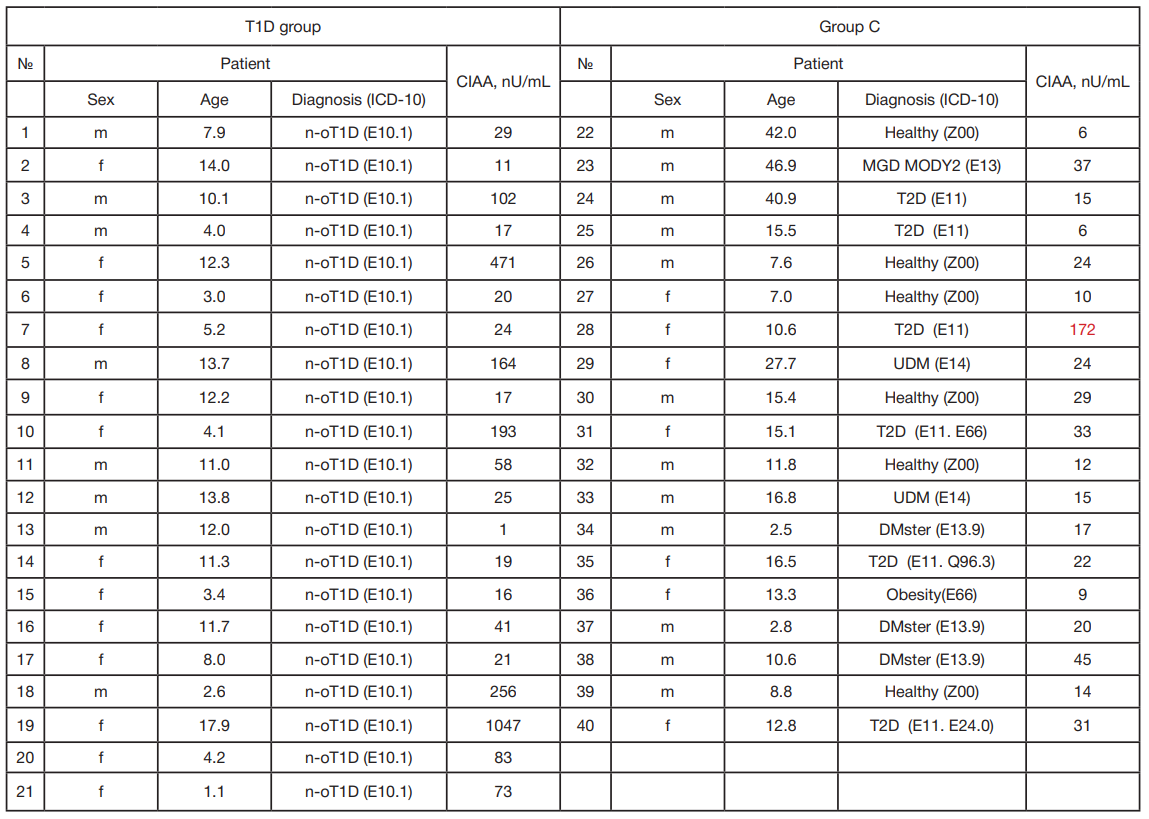
The CIAA measurement results are provided in tab. 2.
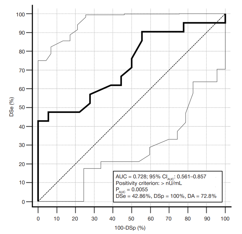
One result (of the serum sample No. 28) was classified as an outlier. Thus, statistical analysis included the results of CIAA measurement in 21 serum samples of the T1D group and 18 serum samples of group C. In the T1D group, the CIAA values varied between 1 and 1047 nU/mL, in group C these varied between 6 and 45 nU/mL. When plotting the test operating characteristic curve, the MedCalc software automatically selected the CIAA value exceeding 45 nU/mL as a test positivity criterion (presence of IAA in the serum sample). DSe, DSp, and DA of the test calculated based on the AUC using the above criterion were 42.9%, 100%, and 72.8% with the 95% confidence intervals 21.8–66%, 81.5–100%, and 56.1–85.7%, respectively (fig. 2).
DISCUSSION
According to IASP data, the median DSe, DSp, and DA of various RIA IAA methods are 44%, 100%, and 81.1 respectively (). DSe of our method (42.9%) is very close to the median DSe of the IASP RIA, and DSp matches the median IASP DSp, but does not fall into its interquartile range. However, DA of our method (72.8%) turned out to be significantly lower compared to the IASP RIA DA and did not fall into its interquartile range.
We explain discrepancy between the result of our RIA method and the RIA results obtained in other IASP labs (the lower DA of our method) by two factors:
- very small number of individuals in both groups;
- the incubation buffer solution (IBS) used by J. Palmer, et al. [4] contained bovine γ-globulin in a concentration of 0.025% that blocked nonspecific insulin binding with the non-IAA immunoglobulins in the serum samples. There was no such reagent in our IBS.
It should be noted that the performance characteristics of our IAA testing methods turned out to be better than that of the ELISA and CLIA methods represented in IASP, and were significantly superior to performance characteristics of the abovementioned Orgentec Anti-Insulin test system popular in Russian labs.
CONCLUSIONS
Ultimately, we regard our results as successful, since the performance characteristics of our method turned out to be much better than that of the ELISA and CLIA methods. We believe that after appropriate adjustment our RIA IAA test can be used for scientific purposes and in clinical practice. Unfortunately, it is currently impossible to use this method in the diagnostic lab of the Russian Children's Clinical Hospital due to two factors: lack of the gamma counter; lack of facilities for radionuclide handling licensed by Rospotrebnadzor and Roszdravnadzor. However, we hope that over time we will manage to tune RIA IAA the Russian diabetologic science and practice are in need of in the Russian Children's Clinical Hospital, branch of the Pirogov University.