ORIGINAL RESEARCH

Two HMG domains of yeast mitochondrial protein Abf2p have different affinity to DNA

Kurashenko AV1, Samoilova EO1, Balaeva MV1, Chicherin IV1, Petrov DYu2, Kamenski P1, Levitskii SA1
About authors

1 Department of Molecular Biology, Faculty of Biology,
Lomonosov Moscow State University, Moscow, Russia

2 Department of General Surgery, Faculty of Fundamental Medicine,
Lomonosov Moscow State University, Moscow, Russia

Correspondence should be addressed: Peter Kamenski
Leninskie gory, d. 1, str. 12, Moscow, Russia, 119991; moc.liamg@iksnemak.rtoip

About paper

Funding: this study was supported by the Russian Foundation for Basic Research (project no. 14-04-31554 mol_a).
Received: 2015-09-29 Accepted: 2015-12-09 Published online: 2017-01-05
|
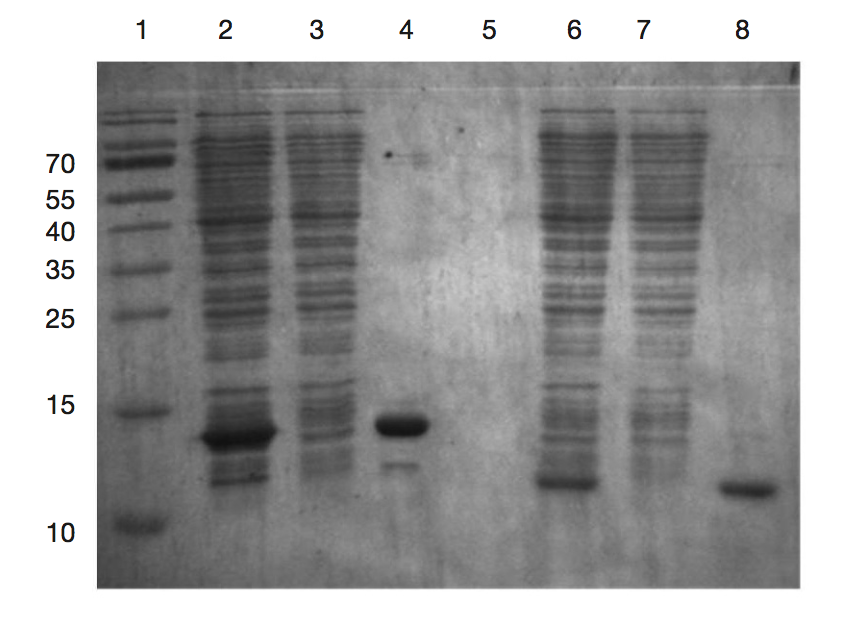
Fig. 1. Isolation and purification of recombinant proteins corresponding to HMG1 and HMG2 domains of the Abf2p protein
Recombinant proteins were purified by metal affinity chromatography on Ni sepharose. 1 — molecular weight markers (the molecular weights of marker proteins are shown on the left); 2, 6 — damaged cell lysates; 3, 7 — factions that didn’t interact with affinity column; 4, 8 — purified preparations of recombinant proteins HMG1 and HMG2 respectively.
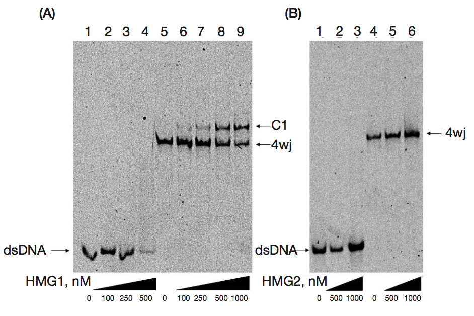
Fig. 2. Analysis of the binding of recombinant proteins corresponding to HMG1 and HMG2 domains with DNA using EMSA
Linear DNA duplex (double-stranded DNA - dsDNA) and cruciform DNA (4-way junction — 4wj) at a concentration of 10 nM were incubated with increasing concentrations of recombinant proteins, after which the reaction mixtures were separated in 6 % polyacrylamide gel. (A) Binding with DNA of HMG1 domain; 1–4 — interaction with DNA duplex, 5–9 — binding with cruciform DNA; C1 — resulting complex. (B) Binding with DNA of HMG2 domain; 1–3 — interaction with DNA duplex, 4–6 — binding with cruciform DNA. The lower part of the figure shows the used concentration of recombinant proteins (in nM).