ISSN Print 2500–1094    ISSN Online 2542–1204
BIOMEDICAL JOURNAL OF PIROGOV UNIVERSITY (MOSCOW, RUSSIA)

New articles

The exploration of ATM gene haplotype evolutionary variability and its geographic dissemination across Eurasia enables the utilization of these data to predict the distribution of diseases associated with alleles comprising these haplotypes. The study aimed to identify the ATM gene haplotypes unique to Eurasia based on the panel of 28 polymorphic loci determining two major “yin-yang” haplotypes. The analysis was conducted using individual genotype data for 28 ATM gene loci across 41 groups of indigenous Eurasian populations. Haplotypes were identified using the Haploview 4.1 software. Phylogenetic analysis was performed in RStudio using the pegas and geneHapR packages. Cartographic visualization of ATM haplotype frequencies was implemented using the GeneGeo cartographic package. The H7j haplotype was identified in indigenous peoples of North Eurasia, which suggests its localized origin. The maximum H7i frequency was reported for Finno-Ugric peoples of the north and indigenous peoples of the northern Eurasia. The highest H8j haplotype frequency is observed in populations of southern China and Japan, which probably indicates the place of its origin. The remaining haplotypes are rare, these are typical for small geographic regions: H6j is sporadically distributed across Asia, H8i is localized to Transcaucasia, and H9i is found in two meta-groups of Eastern European peoples. The data obtained suggest the presence of the ATM gene haplotypes specific for the population of Eurasia, emphasize the contribution of intragenic recombination in shaping the gene haplotype diversity, and highlight the importance of the geographic and demographic context for preservation and spread of distinct genetic variants.
VIEWS 14
Rapid palatal expansion may affect not only maxillary dimensions but also upper airway patency in adolescents; however, the role of tooth-supported and miniscrewassisted appliances remains to be clarified. This study aimed to assess morphological and functional changes in the upper respiratory tract associated with rapid palatal expansion and to compare these changes between the use of Hyrax and MARPE systems. We conducted a prospective pilot study involving 24 adolescents aged 12–16 years with transverse maxillary insufficiency. Fourteen participants received Hyrax appliances, while ten received MARPE devices. Time points: T0 — before treatment, T1 — 6 months after completion of activation. The measured indicators were the volume of the nasal cavity, nasopharynx, and oropharynx, the minimum area of the retropalatal level, and the total nasal resistance at 150 Pa. We also calculated the NOSE score. After treatment, the volume of the nasal cavity increased by 14.6% (p < 0.001), nasopharyngeal volume by 8.9% (p = 0.003), the minimum area of the retropalatal level by 12.1% (p = 0.012), total nasal resistance decreased by 24.7% (p < 0.001), and NOSE score by 35.4 points (p < 0.001). The change in oropharyngeal volume was insignificant (p = 0.091). We established no differences between Hyrax and MARPE groups (p > 0.05). Rapid palatal expansion improved nasal cavity and nasopharynx parameters. Further comparisons of techniques require a priori sample size calculations.
VIEWS 124
High prevalence of postoperative ventral hernias necessitates the development of effective rehabilitation protocols to restore abdominal muscle function early and reduce recurrence risk. This study aimed to evaluate the effects of electromyostimulation (EMS) of the anterior abdominal wall muscles on physical activity levels and strength performance in patients after surgical treatment for postoperative ventral hernias. We enrolled 128 patients (mean age, 47.9 ± 8.6 years) who underwent open combined component separation surgery with retromuscular mesh implantation. In the treatment group (n = 64), the course of anterior abdominal wall EMS (enabled by COMPEX SP-2.0) started on the 10th day after surgery; in the control group (n = 64), we followed a standard post-surgery protocol. The assessed indicators were the level of physical activity (pedometry, 7 days) and the strength of the trunk muscles (strain dynamometry, registered with a BackCheck 700 device). Six months after surgery, the EMS group showed significantly greater muscle strength gains than the control group: in trunk extension, median strength was 26.9 kg versus 15.4 kg (74.7% increase; p < 0.001); in flexion, it was 15.7 kg versus 8.0 kg (96.3% increase; p < 0.001). The level of physical activity in the EMS group was significantly higher: the median number of steps per week was 27304.5 ± 2903.48 (95% CI 20964.6–33644.5), while in the control group it reached 11173.6 ± 3688.8 (95% CI 10065.4–12281.9) steps. This indicates a recovery to 90.0% of the preoperative level. Thus, an early post-surgery course of EMS of the anterior abdominal wall muscles is an effective rehabilitation method that significantly improves the strength characteristics of the core muscles and restores the levels of physical activity in patients.
VIEWS 150
Muscular dysfunction of the anterior abdominal wall persists in many patients post component separation due to postoperative ventral hernia. Electrical myostimulation can contribute to better recovery, but its efficacy after such surgical procedures is poorly understood. The study aimed to assess the effect of the postoperative electrical myostimulation on the neuromuscular conduction and functional activity of the rectus abdominis muscles. A total of 128 patients (average age 47.9 ± 8.6 years) post component separation were included in a prospective controlled non-randomized study. The index group (n = 64) received electrical myostimulation starting from day 10 (12 sessions, 5–10 min each, 3 times a week, COMPEX SP-2.0® muscle stimulator, Switzerland), and the control one (n = 64) received no electrical myostimulation. Electroneuromyography of the rectus abdominis muscles was performed before and after the course using the Synapsis system (Neurotech, Russia). In the index group, the latency period reduced from 10.1 to 7.9 ms (by 21.8%; p < 0.001), and in the control group it reduced from 9.7 to 9.2 ms (by 5.2%; p < 0.001); the intergroup difference p = 0.002. The M-response amplitude improved in both groups (index group: from 8.4 to 8.9 mV, +5.6%, p < 0.001; control group: from 8.2 to 8.8 mV, +6.8%, p < 0.001), without any intergroup differences (p = 0.295). The induced muscle contraction velocity changed minimally in the index group (from 45.0 to 45.4 m/s, p = 0.049) and did not change in the control group (p = 0.316); in 89.1% of patients, the values were still below normal. Conclusions: postoperative electrical myostimulation significantly accelerates the neuromuscular conduction restoration, but does not affect the muscular response amplitude. It is reasonable to include electrical myostimulation in rehabilitation programmes.
VIEWS 230

Popular articles

The use of allogenic bone material as a ceramic filler for DLP printing makes it possible to obtain personalized complex-shaped implants combining the matrix biomimetic nature with the additive technology benefits. The study aimed to assess the possibility of using the calcined cortical bone allograft powder as part of photopolymerizable suspension for DLP printing and producing bioceramics with the characteristics comparable to that of synthetic hydroxyapatite by sintering. The bone allograft was subjected to multi-stage specialized treatment involving complete removal of cells with preservation of the intercellular matrix and collagen fiber structure. The calcined medical allograft was crushed, introduced into a photopolymerizable matrix, and used for DLP printing of the samples that were further sintered and analyzed by X-ray diffraction and energy-dispersive spectroscopy methods before and after additive production. The sintered material specific gravity was 81.5%, compressive strength — 75.8 MPa, tensile strength — 12 MPa, Young's modulus — 3.08 GPa, and Vickers hardness — 0.55 GPa, which was within the range of values for porous hydroxyapatite. After DLP printing and sintering the sample phase and elemental composition did not change considerably compared to the source calcined material. The calcined bone allograft powder is suitable for preparing photopolymerizable suspensions and subsequent DLP printing of ceramic samples without deteriorating the material phase and chemical stability. The resulting mechanical properties make it possible to consider this allogenic bone material as a promising candidate for production of personalized implants with sophisticated geometry.
VIEWS 1243
Monoclonal antibody therapy is one of the most promising approaches for effective influenza control. In this study, we evaluated the antiviral activity of exogenous mRNA-encoded single-chain variable fragment (scFv) antibodies, which are capable of binding viral antigens inside the cell with high affinity. Two influenza virus proteins, hemagglutinin (antibody FI6) and nucleoprotein (antibody 2/3), were chosen as targets. Each scFv encoded by mRNA was produced in two variants: one containing a signal peptide (SP) to direct secretion into the extracellular space (scFv-SP) and one lacking the signal peptide (scFv-WO) for cytosolic localization and function. These variants showed distinct intracellular localization patterns: scFv-SP localized to regions characteristic of the endoplasmic reticulum and the Golgi complex, whereas scFv-WO was distributed diffusely throughout the cytoplasm. mRNAs encoding scFv-FI6-SP, scFv-2/3-SP, and scFv-2/3-WO exhibited antiviral activity against influenza A virus in vitro. The scFv-FI6-SP mRNA showed the strongest antiviral effect, reducing viral load by approximately tenfold compared to the control. For influenza B virus, both  scFv-2/3 mRNA variants, with and without the signal peptide,  reduced viral load by an average of 50%. These findings highlight the antiviral potential of intracellular antibodies and point to new opportunities for targeting viral components that are not accessible to conventional antiviral therapies.
VIEWS 1112
Dear researcher!
At the end of 2015, Bulletin of RSMU saw an important change in its typographic design and content. We formulated new editorial policies and established strict ethical standards for submitted manuscripts in accordance with the guidelines of reputable international bodies. As a result, about a quarter of the submitted works have been rejected, the primary reason being the author trying to submit a previously published article. Sometimes authors believe that by making slight changes to the introduction, excluding a few people from the study, performing a new statistical analysis, and thus obtaining totally new results they will turn their old manuscript into a novel work. That is why we would like to talk about scientific integrity, honesty, plagiarism, and self-plagiarism in our special project “Author’s work”.
Richard FEYNMAN Cargo cult science
American physicist Richard P. Feynman, a Nobel laureate, was always very scrupulous about the quality of a research study. During his commencement address at the California Institute of Technology in 1974, he talked about scientific integrity and honesty and warned young researchers “not to fool” themselves. A must-read for anyone who believes he/she is a true scientist.
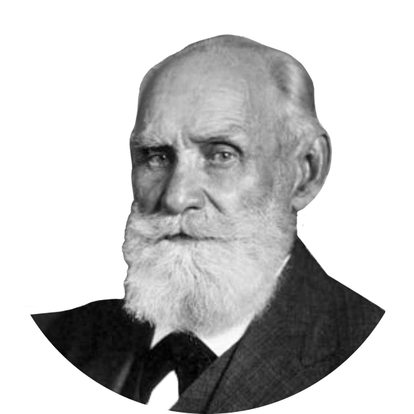
Ivan PAVLOV On the Russian mind
In 1918, Russian physiologist Ivan Pavlov, a Nobel laureate, delivered two lectures: on the mind in general and the Russian mind in particular; on those mind qualities that determine the success of a research work and on how these qualities are present in the Russian mind. Pavlov's thoughts are an effective vaccine against poor intellectual work.