Copyright: © 2017 by the authors. Licensee: Pirogov University.
This article is an open access article distributed under the terms and conditions of the Creative Commons Attribution license (CC BY).

ORIGINAL RESEARCH

Study of ablated surface smoothness and thermal processes in rabbit cornea treated with MicroScan-Visum and MicroScan-PIC excimer laser systems

About authors

Department for Clinical Research in Ophthalmology,
Clinical Research Center for Otorhinolaryngology, Moscow, Russia

Correspondence should be addressed: Nika Takhchidi
Volokolamskoe shosse, d. 30, str. 2, Moscow, Russia, 123182; ur.ay@ht-akin
Received: 2016-04-11 Accepted: 2016-04-14 Published online: 2017-01-05
|
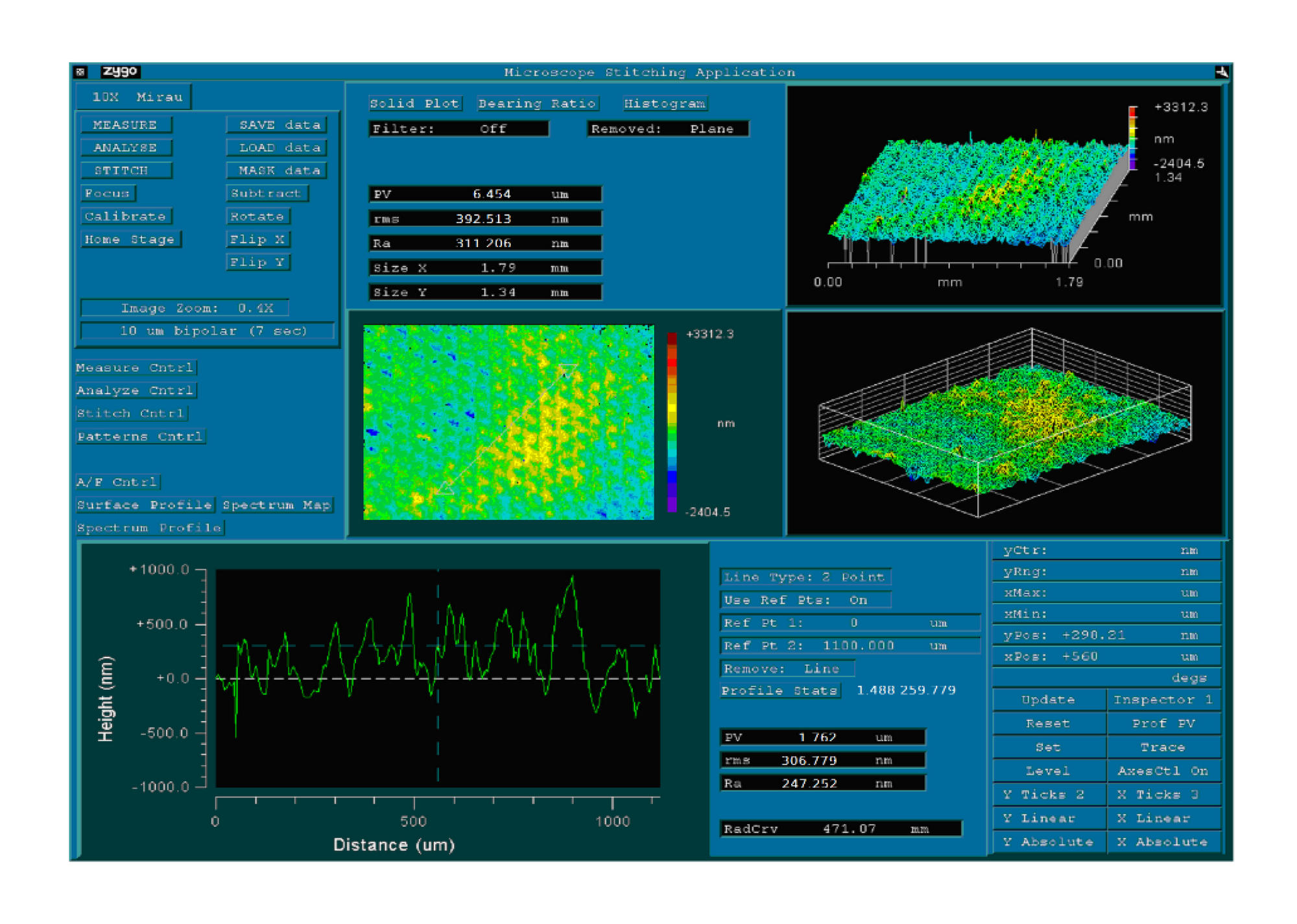
Fig. 1. Example of measurement of the smoothness of the ablation surface formed using MicroScan-PIC 100 Hz by interferometer New View – 5000 Zygo (Ra = 311 nm)
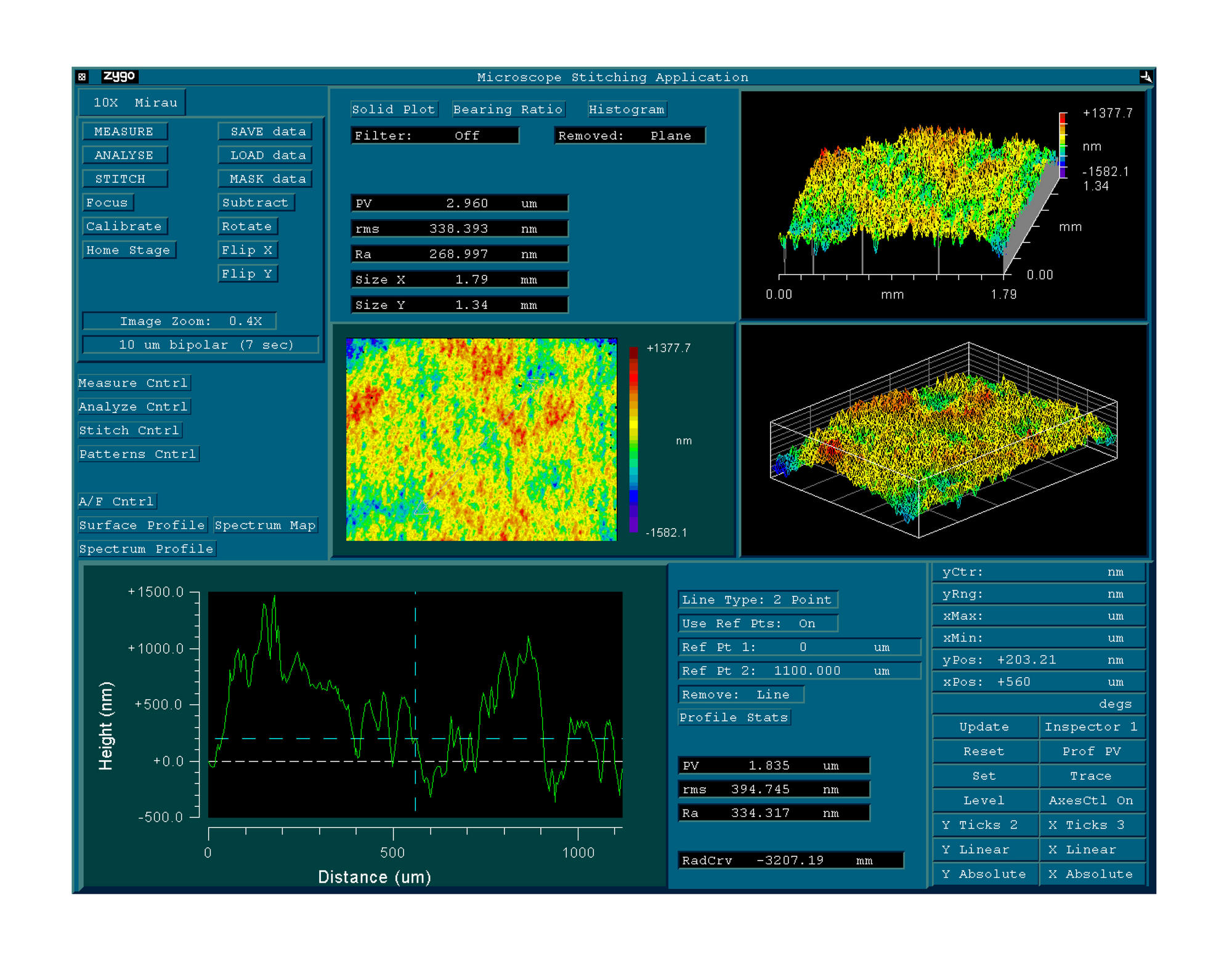
Fig. 2. Example of measurement of the smoothness of the ablation surface formed using MicroScan-Visum 500 Hz by interferometer New View – 5000 Zygo (Ra = 268 nm)
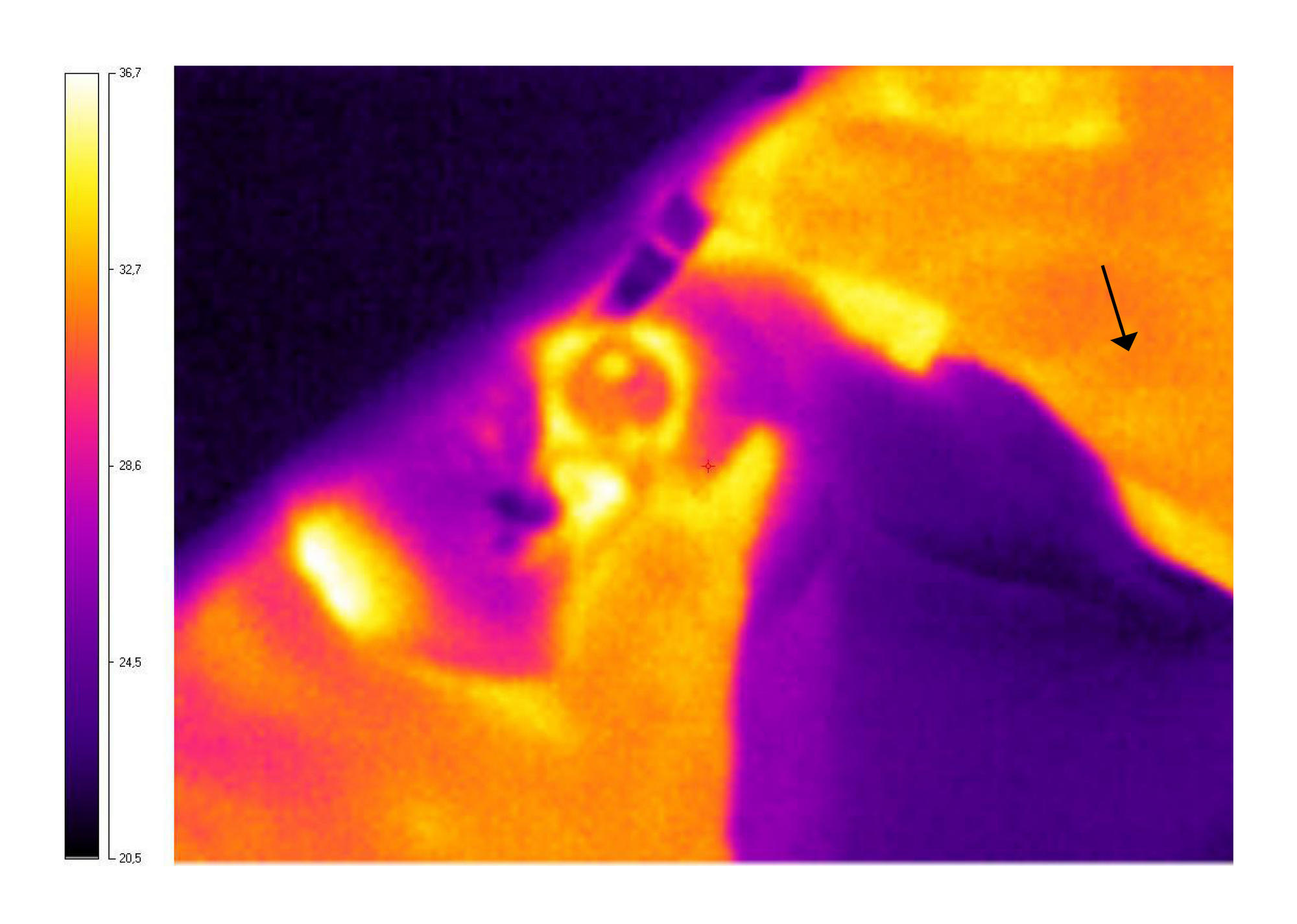
Fig. 3. Example of thermographic map during phototherapeutic keratectomy using MicroScan-PIC 100 Hz (rabbit cornea is marked by an arrow)
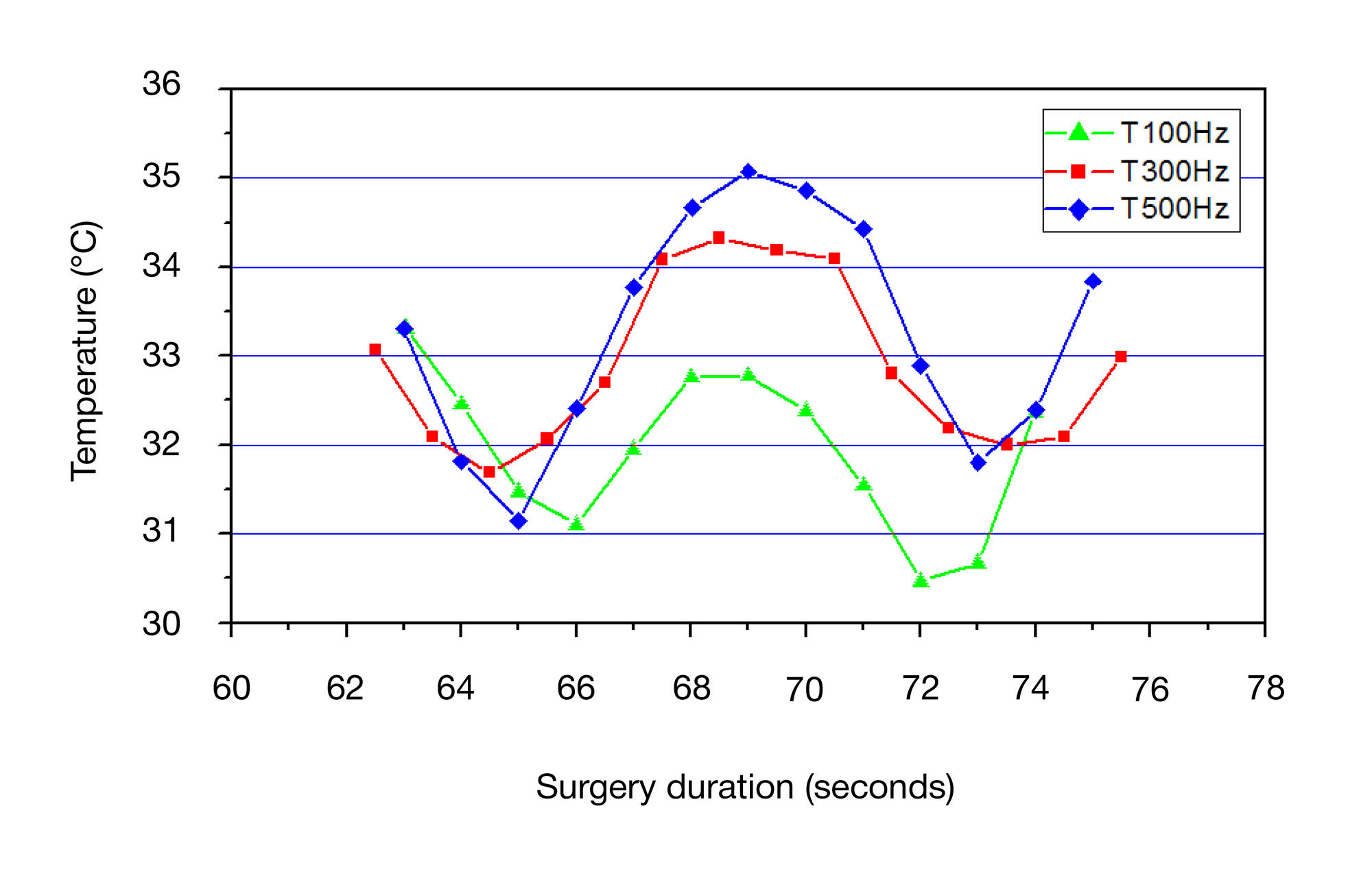
Fig. 4. Transverse temperature profiles for excimer laser systems MicroScan- PIC 100 Hz (green curve), MicroScan-Visum 300 Hz (red curve) and MicroScan-Visum 500 Hz (blue curve)
Table 1. Indicators of the ablation surface quality of lens formed on polymethylmethacrylate plates using radiation from MicroScan-PIC 100 Hz, MicroScan-Visum 300 Hz and MicroScan-Visum 500 Hz systems, nm (M ± δ, p < 0.05)
Table 2. Dynamics of thermal processes in a rabbit cornea during phototherapeutic keratectomy using laser systems MicroScan-PIC 100 Hz, MicroScan-Visum 300 Hz and MicroScan-Visum 500 Hz, °C (M ± δ, p < 0.05)