Copyright: © 2017 by the authors. Licensee: Pirogov University.
This article is an open access article distributed under the terms and conditions of the Creative Commons Attribution license (CC BY).

ORIGINAL RESEARCH

Postmortem magnetic resonance imaging in the diagnosis of congenital pneumonia

Tumanova UN1, Lyapin VM1, Bychenko VG1, Schegolev AI1,2, Sukhikh GT1
About authors

1 Kulakov Research Center for Obstetrics, Gynecology and Perinatology, Moscow, Russia

2 Pirogov Russian National Research Medical University, Moscow, Russia

Correspondence should be addressed: Uliana Tumanova
ul. Akademika Oparina, d. 4, Moscow, Russia, 117997; moc.liamg@avonamut.n.u
Received: 2016-08-17 Accepted: 2016-08-23 Published online: 2017-01-05
|
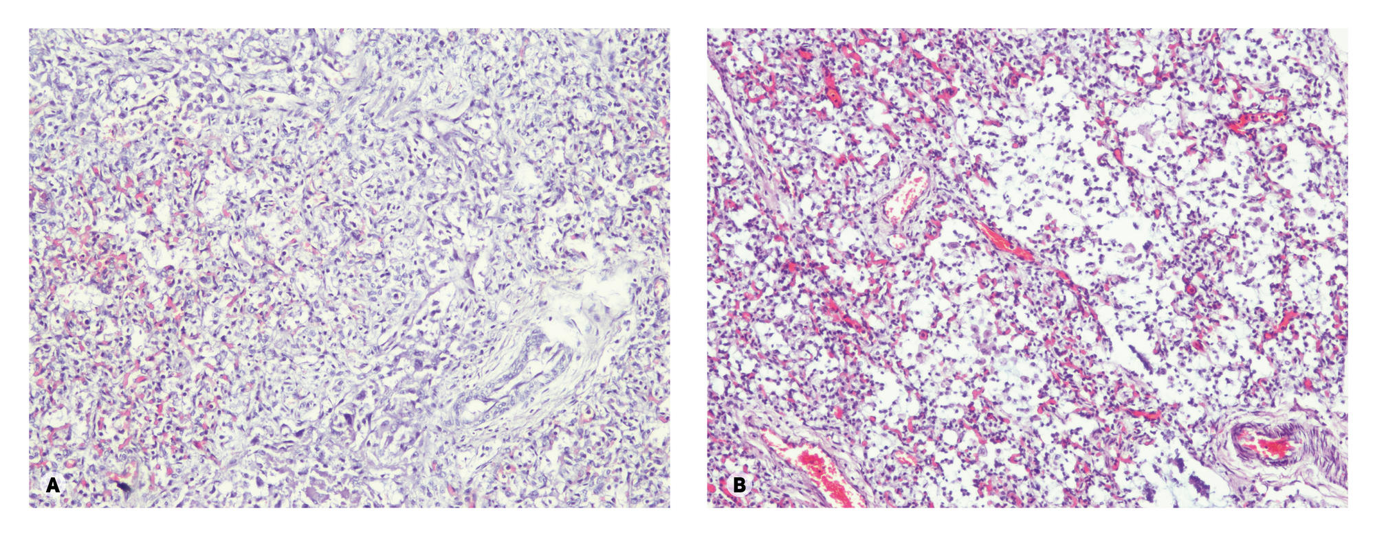
Fig. 1. Histological changes of lungs: (A) hypoplasia, (B) monocyte/macrophage pneumonia. Staining with hematoxylin and eosin, ×100
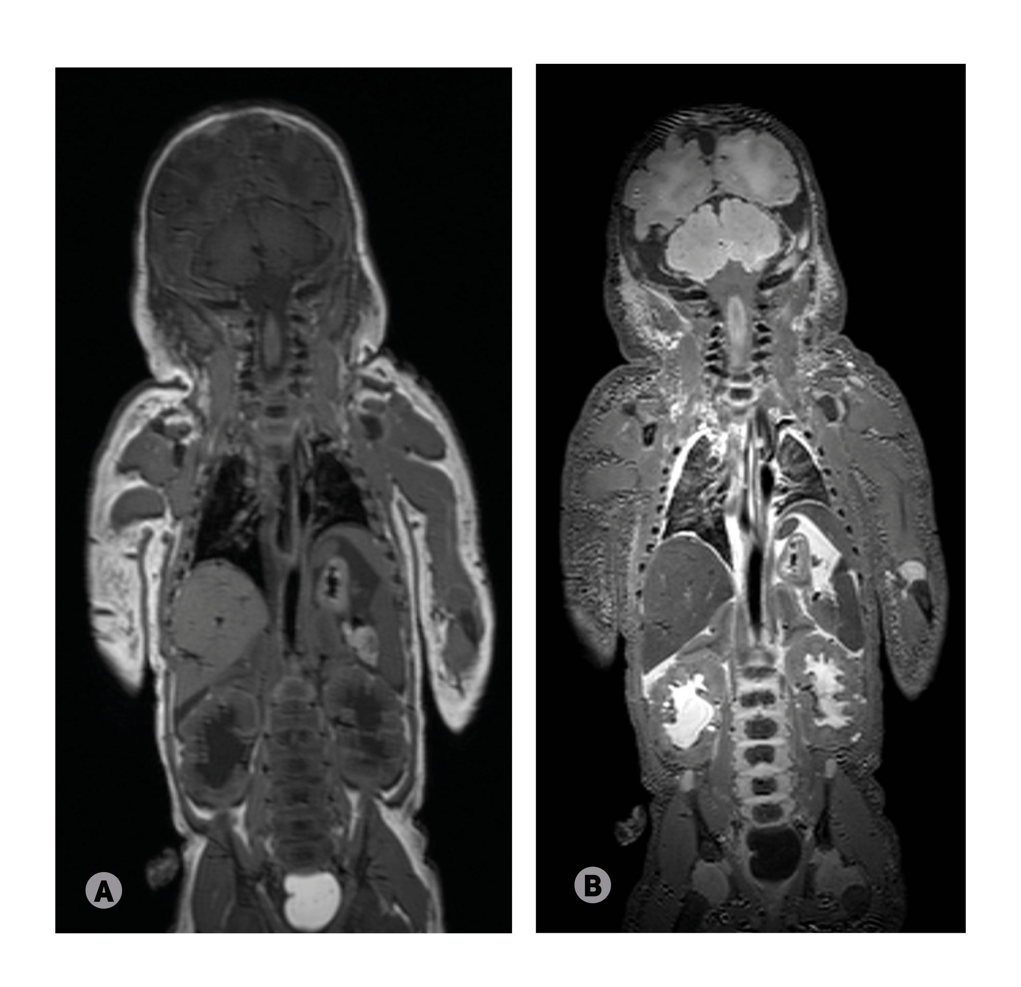
Fig. 2. Coronal MR-images of a dead neonate with congenital pneumonia (the experimental group): (A) T1-weighted image, (B) T2-weighted image
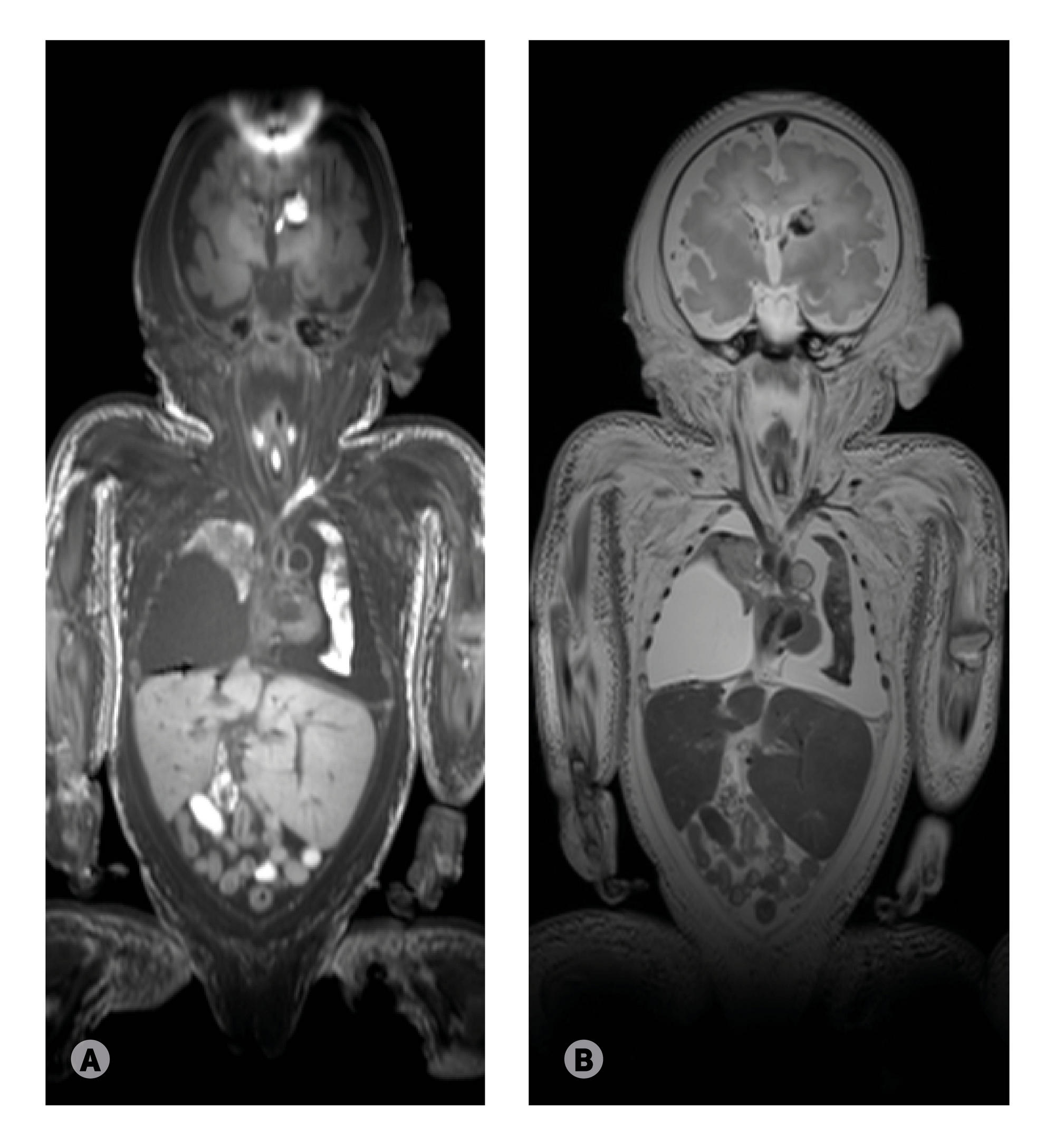
Fig. 3. Coronal MR-images of a dead neonate without lung pathology (the comparison group): (A) T1-weighted image, (B) T2-weighted image
Fig. 4. Coronal MR-images of a dead neonate from the comparison group with hemorrhages in both lungs in the absence of pneumonia: (A) T1-weighted image, (B) T2-weighted image
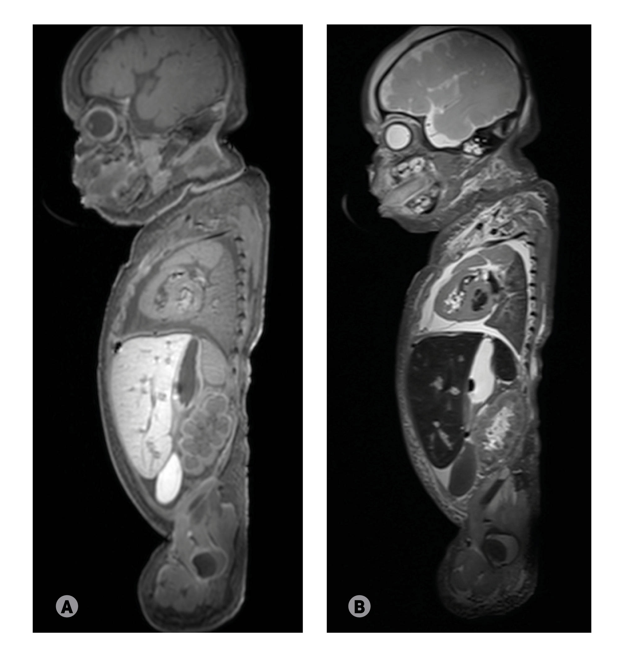
Fig. 5. Sagittal MR-images of a dead neonate from the comparison group with the intact left lung and right lung compressed by a diaphragmatic hernia
Table 1. Clinical and morphological features of dead neonates
Table 2. Signal intensity and airiness index (AI) of lungs of dead neonates