ORIGINAL RESEARCH

Nuclear medicine in the diagnosis of renal and coronary angiopathies in patients with type 2 diabetes and impaired glucose tolerance

Kharina DS1,2, Poletaeva TA1, Kondakov AK1,2, Mosin DYu1, Znamensky IA1,2
About authors

1 Central Clinical Hospital of RAS, Moscow, Russia

2 Pirogov Russian National Research Medical University, Moscow, Russia

Correspondence should be addressed: Dina Kharina
Litovsky bulvar, d. 1a, Moscow, Russia, 117593; ur.liam@ls.anidras
Received: 2016-11-25 Accepted: 2016-12-10 Published online: 2017-01-19
|
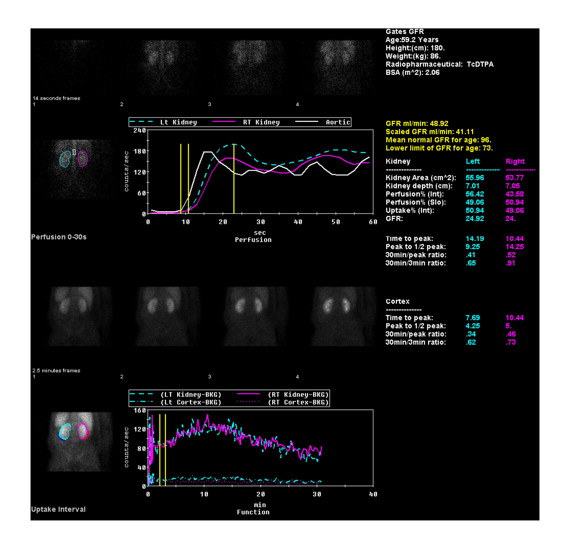
Fig. 1. Reconstruction of kidney images obtained by dynamic renal scintigraphy using Xeleris 2.1 working station (General Electric, USA) and Renal Analysis software (Emory University Hospital, USA)
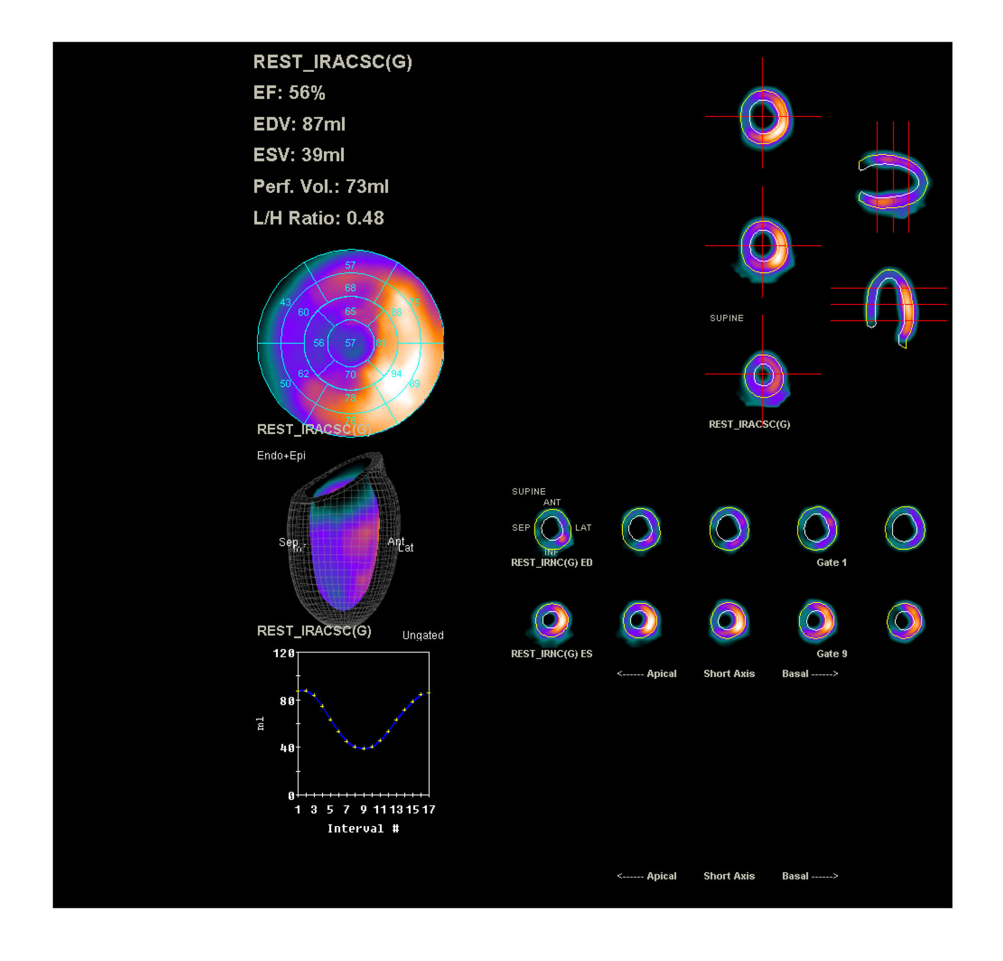
Fig. 2. Reconstruction of myocardium images obtained by perfusion SPECT using Quantitative Gated SPECT/Quantitative Perfusion SPECT software (Cedars-Sinai Hospital, USA)
Table 1. Demographic characteristics of patient subgroups based on the radionuclide diagnostic technique applied
Abbreviations: type 2 DM — type 2 diabetes mellitus; IGT — impaired glucose tolerance; M — arithmetic mean; SD — standard deviation.
Table 2. Glomerular filtration rates calculated using Gates method (dynamic renal scintigraphy) and Cockroft–Gault equation
Abbreviations: type 2 DM — type 2 diabetes mellitus; IGT — impaired glucose tolerance. Data are presented as M ± SD (μ), where M is arithmetic mean, SD is standard deviation, μ is median.
Table 3. Results of blood biochemistry tests and myocardial perfusion SPECT scans
Abbreviations: type 2 DM — type 2 diabetes mellitus; IGT — impaired glucose tolerance. Data are presented as M ± SD (μ), where M is arithmetic mean, SD is standard deviation, μ is median.