ORIGINAL RESEARCH

Clinical and laboratory features of hemostatic disorders in patients with retinal vein occlusion

Shelkovnikova TV1,2, Takhchidi KhP3, Volkov AN4, Shishlyannikova NYu
About authors

1 Novokuznetsk State Institute of Further Training of Physicians, Novokuznetsk, Russia

2 Kemerovo Regional Clinical Ophthalmological Hospital, Kemerovo, Russia

3 Pirogov Russian National Research Medical University, Moscow, Russia

4 Kemerovo Regional Clinical Hospital, Kemerovo, Russia

5 Kemerovo State Medical University, Kemerovo, Russia

Correspondence should be addressed: Tatiana Shelkovnikova
Pionerskiy bulvar, d. 10a, kv. 1, Kemerovo, Russia, 650066; moc.liamg@avokinvoklehs.t
Received: 2016-11-28 Accepted: 2016-12-10 Published online: 2017-01-19
|
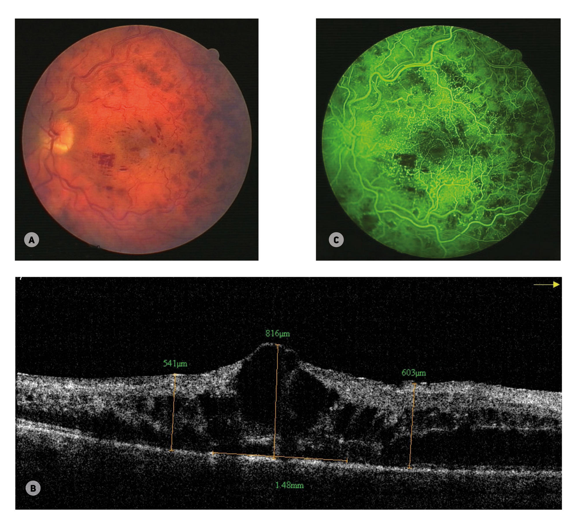
Fig. 1. Patient S., 38 years old. Diagnosed with central retinal vein occlusion; lupus anticoagulant and FV Leiden detected. (A) Conspicuous retinal hemorrhage syndrome. (B) Results of optical cogerence tomography. Diffuse cystoid macular edema; foveal thickness of 816 μm, peri- and parafoveal thickness of 541–603 μm; destroyed pigment epithelium and photoreceptor layer, d = 148 mm. (C) Fundus fluorescein angiography. Leakage from retinal vessels, cystoid macular edema, retinal ischemia
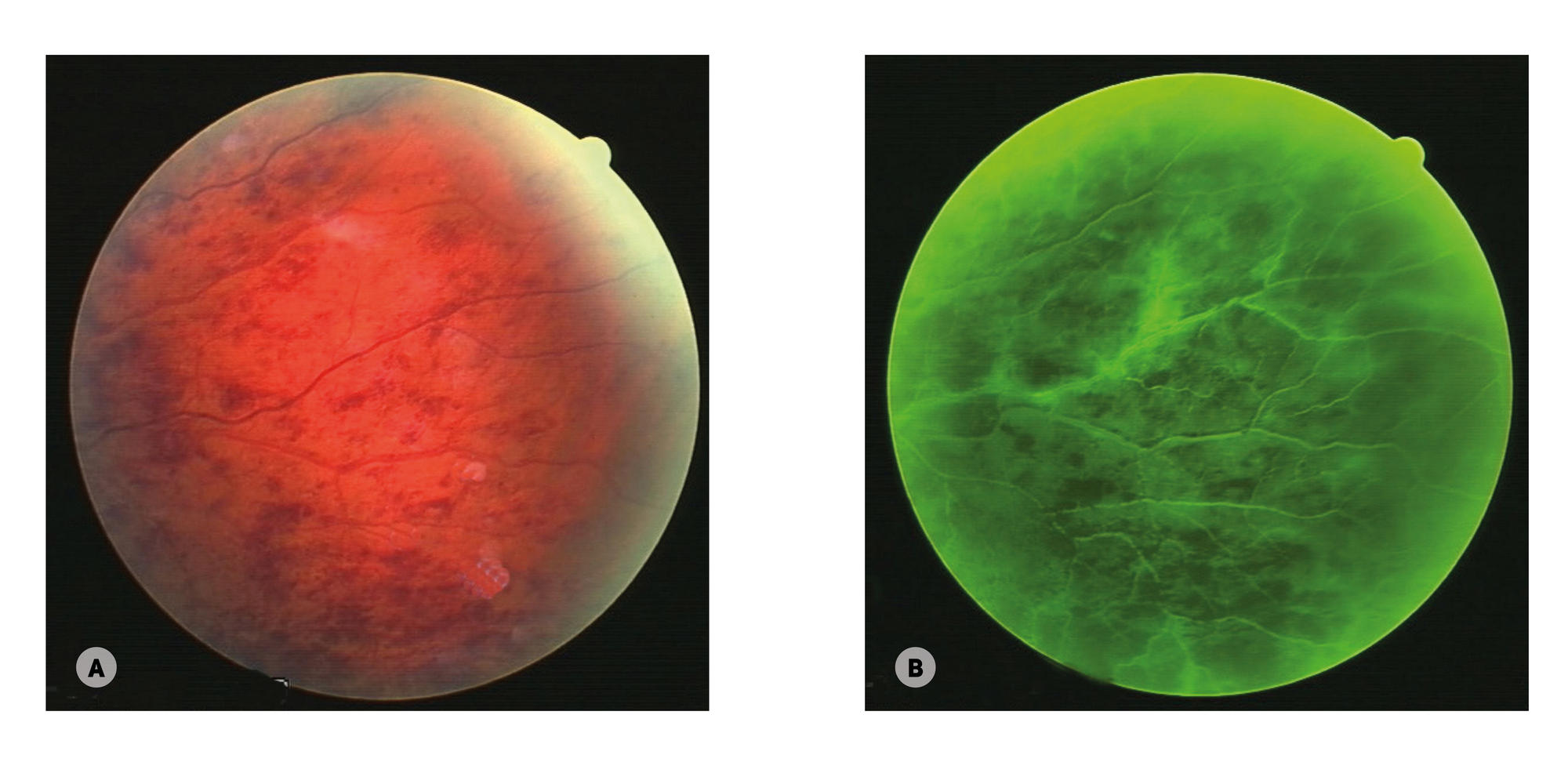
Fig. 2. Patient S., 38 years old. Diagnosed with central retinal vein occlusion; lupus anticoagulant and FV Leiden detected. (A) Medium periphery. Conspicuous retinal hemorrhage syndrome. (B) Fundus fluorescein angiography. Leakage from retinal vessels, cystoid macular edema, retinal ischemia
Homeostasis in patients with retinal vein occlusion depending on the presence of FV Leiden and lupus anticoagulant (LA) in patients’ blood
a — p < 0.05; comparison of groups 1, 2 and 3 with the controls;
b — p < 0.05; group 1 compared to group 2;
c — p < 0.05 groups 1 and 2 compared to group 3.