ORIGINAL RESEARCH

RNA interference targeting interstitial collagenase is a potential therapeutic tool to treat psoriasis

Mogulevtseva YuA1, Mezentsev AV2, Bruskin SA2
About authors

1 Russian State Agrarian University – Timiryazev Moscow Agricultural Academy, Moscow, Russia

2 Laboratory of Functional Genomics, Department of Genetics and Biotechnology,
Vavilov Institute of General Genetics of the Russian Academy of Sciences, Moscow, Russia

Correspondence should be addressed: Aleksandr Mezentsev
ul. Gubkina, d. 3, Moscow, Russia, 119333; ur.ggiv@vestnesem

About paper

Acknowledgements: the authors thank Prof. Eleonora Piruzian of Vavilov Institute of General Genetics for a critical analysis of the manuscript and Prof. Maria Lagarkova of Vavilov Institute of General Genetics for the plasmids used in this study.

Contribution of all authors in data collection, analysis and interpretation was equal. Mogulevtseva AYu prepared all illustartions and Mezentsev AV drafted a manuscript. All authors participated in editing of the manuscript.
Received: 2017-02-17 Accepted: 2017-03-25 Published online: 2017-07-19
|
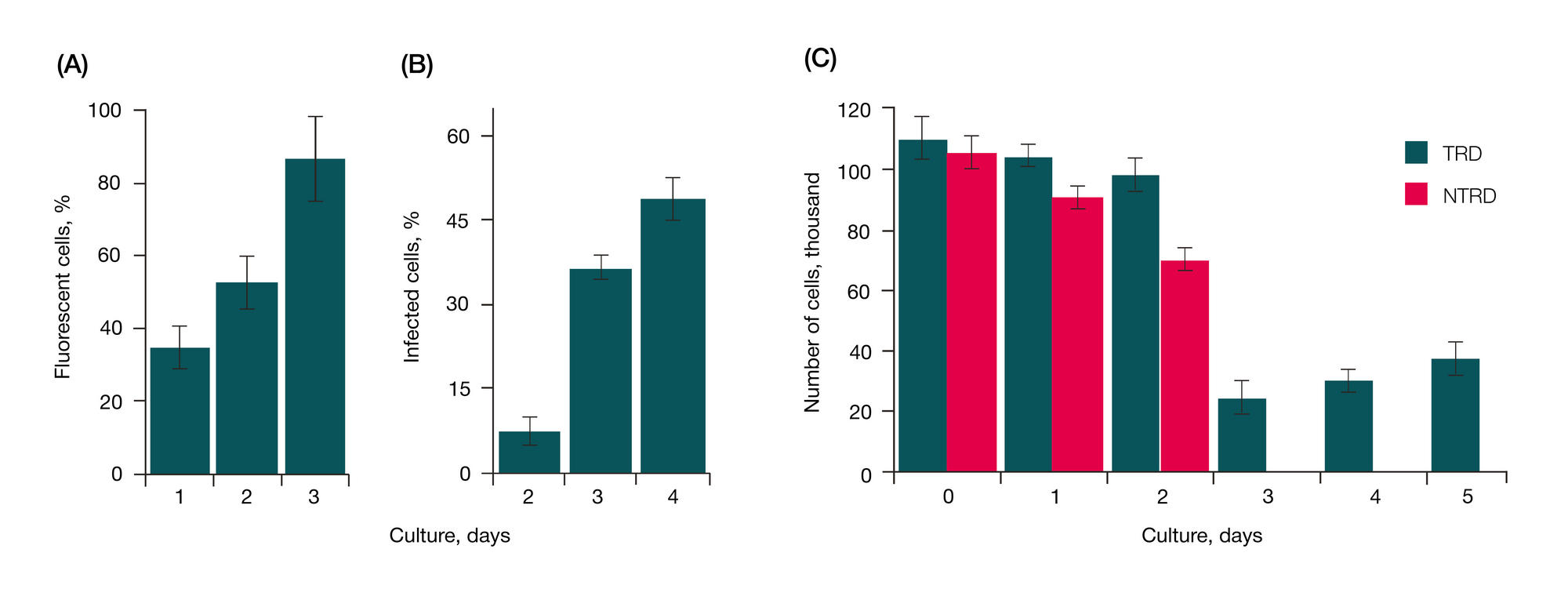
Fig. 1. Lentiviral transduction. (A) Ratio of HEK293 cells emitting the fluorescent light in the course of transfection. (B) Ratio of HaCaT cells emitting the fluorescent light in the course of transduction. (C) Number of viable HaCaT cultured in the presence of puromycin. TRD — transduced cells; NTRD — non-transduced cells (negative control). Transfection and transduction were performed as described in Methods
Fig. 2. Effect of mRNA expression on the morphological characteristics of transduced cells. (A) Cells expressing control shRNA. (B) Cells expressing shRNA specific to interstitial collagenase. Transduction of HaCaT cells was performed as described in Methods
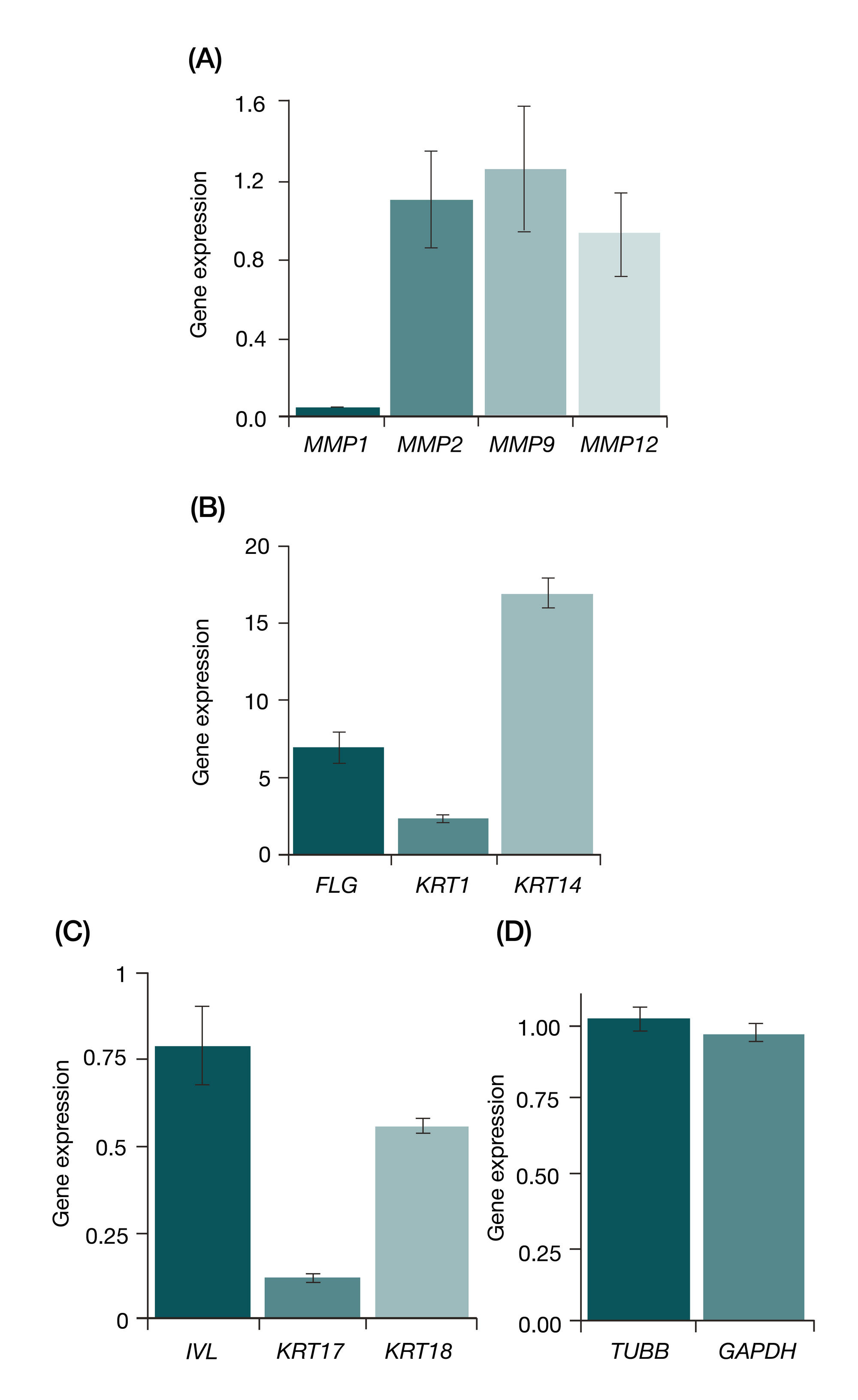
Fig. 3. Assessment of gene expression in transduced human epidermal keratinocytes by quantitative PCR. (A) Shifts in matrix metalloproteinase expression. (B) and (C) Shifts in over- and underexpressed genes. (D) Housekeeping gene expression. Data were normalized to the expression of ACTB. The figure shows results of comparison of gene expression in epidermal keratinocytes expressing interstitial collagenase-specific shRNA and control shRNA (see Methods)
Fig. 4. Effect of expression of in interstitial collagenase-specific shRNA on proliferation and migration of transduced cells. Analysis of cell growth curves (A) and quantitative assessment of migration rate (B) of HaCaT cells expressing IC-specific shRNA and scramble shRNA. (C) Migration of transduced cells: photos taken the next day after transduction began and after it was finished. For details please refer to Methods
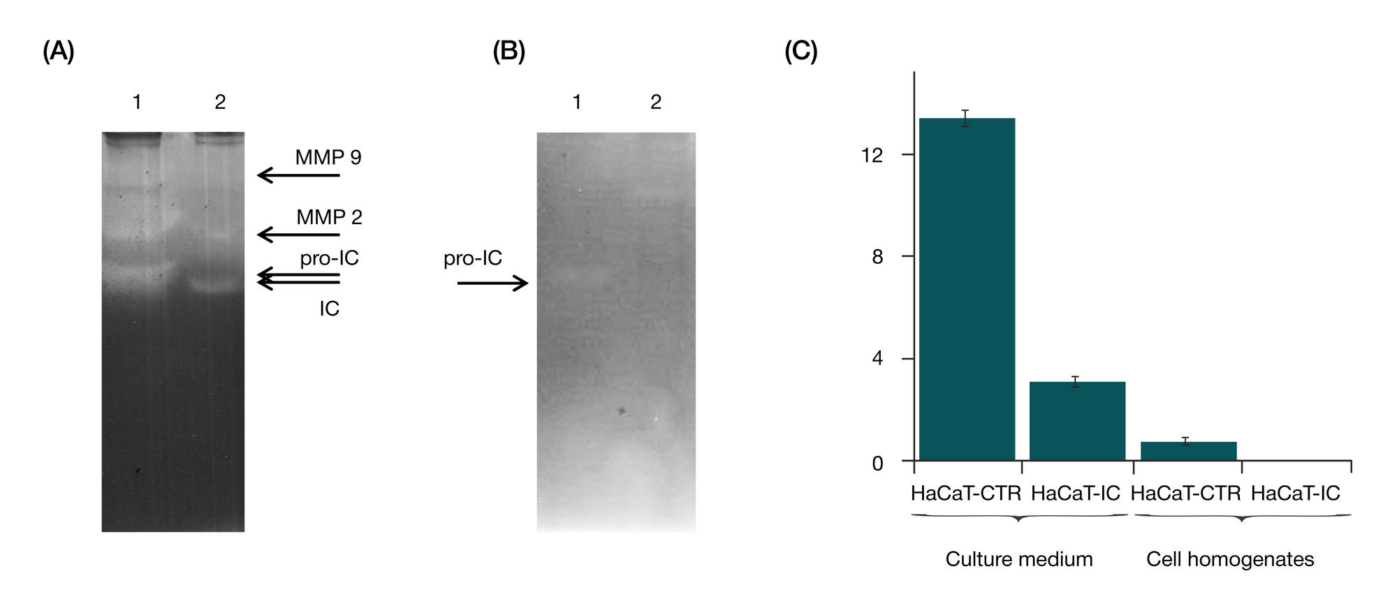
Fig. 5. Analysis of interstitial collagenase activity in transduced epidermal keratinocytes, cell homogenates and culture medium. (A) Zymography of culture medium samples collected from growing transduced cells: 1 — cells expressing scramble shRNA. 2 — cells expressing IC-specific shRNA. The arrows show positions of matrix metalloproteinases in the gel. (B) Zymography of homogenate samples obtained from growing transduced cells: 1 — cells expressing scramble shRNA. 2 — cells expressing IC-specific shRNA. (C) Quantitative analysis of enzymatic activity. Samples were collected 48 hours after culture onset. Confluency at the time of sample collection — 60 %. Cell culturing, preparation of homogenates, details of protein electrophoresis and zymography are described in Methods