METHOD

Knee osteochondritis desiccans: surgery algorithm

Egiazaryan KA, Lazishvili GD, Hramenkova IV, Shpak MA, Badriev DA
About authors

Department of Traumatology, Orthopedics and Field Surgery, Faculty of Pediatrics,
Pirogov Russian National Research Medical University, Moscow

Correspondence should be addressed: Guram D. Lazishvili
ul. Ostrovityanova, 1, Moscow, 117997; 89166575996; moc.liamg@zalmarug
Received: 2018-05-16 Accepted: 2018-06-23 Published online: 2018-06-27
|
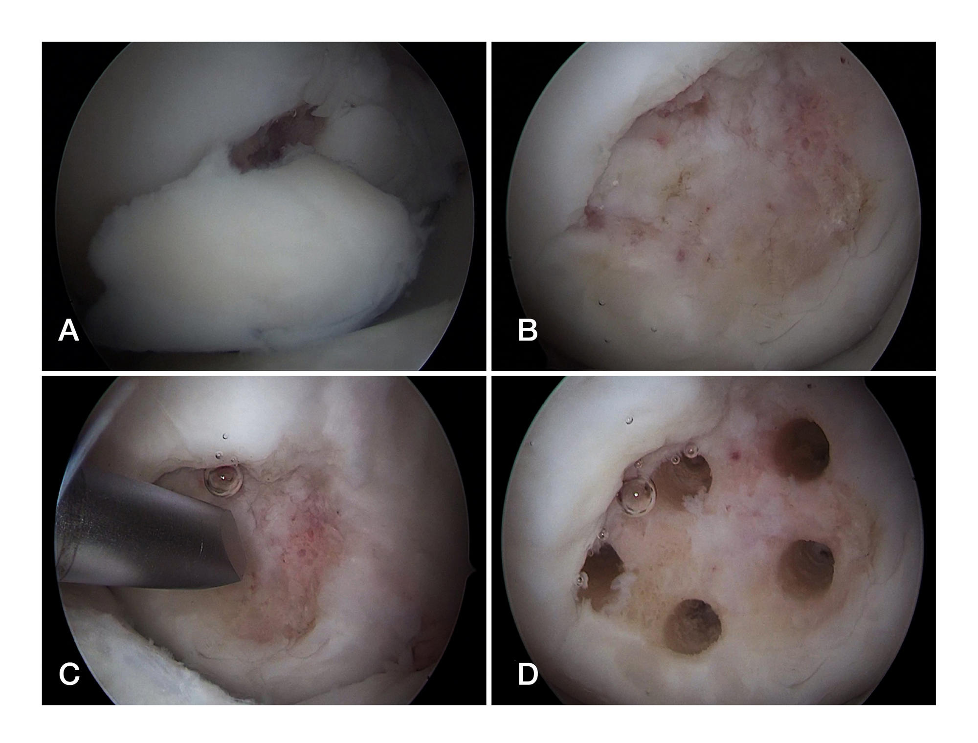
Fig. 1.А. Separated necrotic cartilage plate, arthroscopic view. B. Osteochondral defect after removal of the necrotic plate, arthroscopic view. C. Drilling holes (tunnels) in the subchondral bone. D. Subchondral bone after tunneling
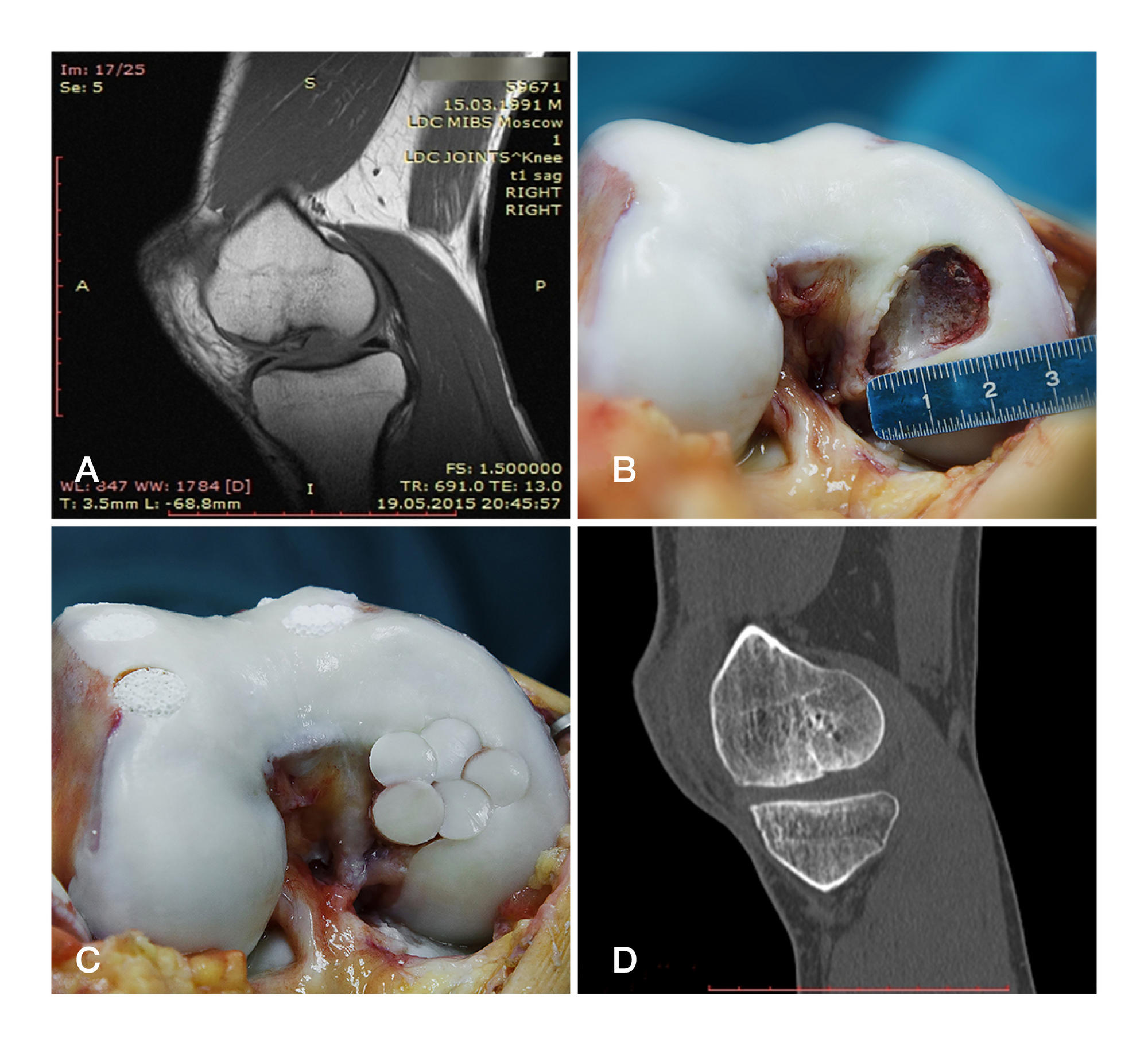
Fig. 2. А. Osteochondritis desiccans affecting medial femoral condyle, MRI scan. B. Condyle defect shape and size after sanation. C. Result of 'mosaic' osteochondral autotransplantation. Donor sites filled with biocomposite bone. D.12 months after operation, MSCT scan. Osteochondral regeneration in the 'mosaic' zone went well
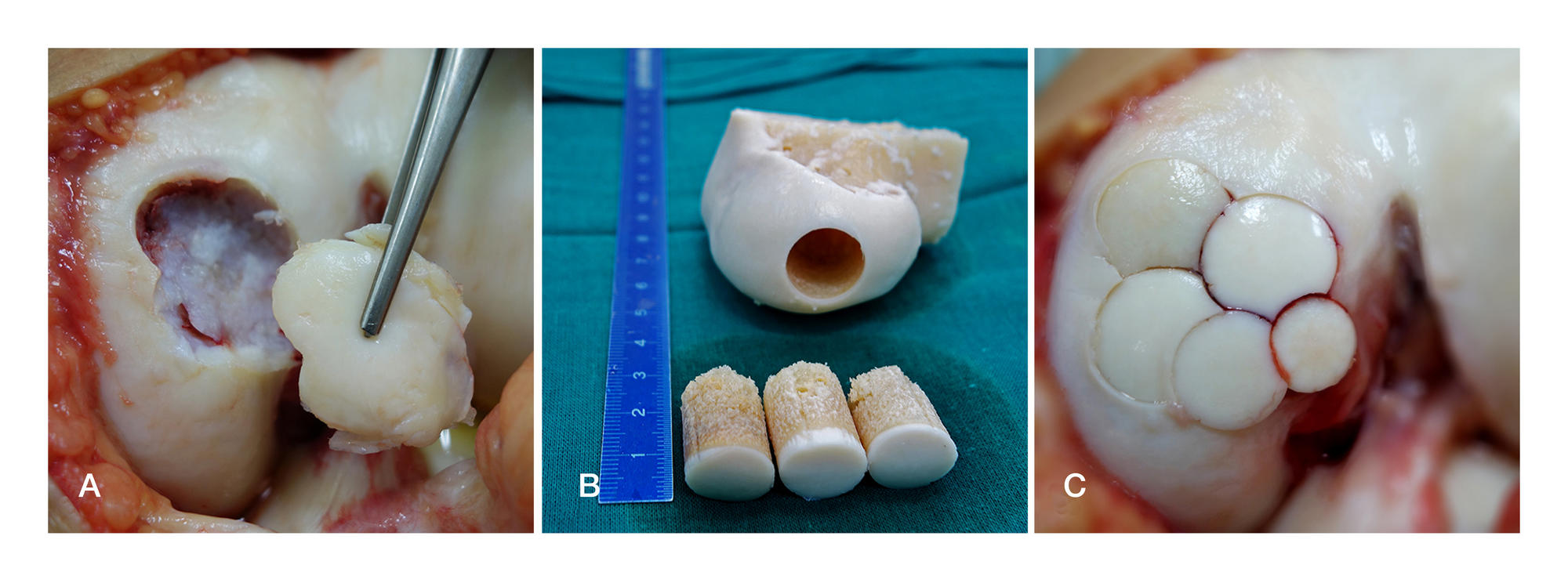
Fig. 3. A. Osteochondral defect of the medial femoral condyle (Koenig's disease). B. Allogeneic lyophilized femoral condyle and cylindrical donor transplants (columns, posts). C. Result of "mosaic" osteochondral allotransplantation
Fig. 4. А. Osteochondral defect of the medial femoral condyle (Koenig's disease), shape and size. B. Result of combined osteochondral autotransplantation. A — auto, E — allo
Fig. 5. А. Osteochondral defect of the medial femoral condyle shape and size after sanation. B. Subchondral bone after tunneling. C. Collagen matrix implantation. D. 1.4 years after surgery: condyle defect is completely covered by stable cartilaginous tissue (arthroscopic view)