Copyright: © 2018 by the authors. Licensee: Pirogov University.
This article is an open access article distributed under the terms and conditions of the Creative Commons Attribution license (CC BY).

ORIGINAL RESEARCH

The study of morphological and functional changes in the thyroid follicles of healthy rats and rats with experimentally induced hypothyroidism following exposure to medium-power laser radiation

About authors

1 Multi-specialty Center of Laser Medicine, Chelyabinsk

2 South Ural State Medical University, Chelyabinsk

Correspondence should be addressed: Irina V. Smelova
Potemkina 14, kv. 65, Chelyabinsk, 454081; ur.liam@vis.larips

Received: 2018-02-15 Accepted: 2018-03-20 Published online: 2018-07-28
|
Fig. 1. The intact thyroid gland. Staining: hematoxylin-eosin; 400x magnification
Fig. 2. Hypothyroidism. Staining: hematoxylin-eosin; 400x magnification
Fig. 3. The thyroid of experimental animals on day 7 after irradiation with 112 J/cm2 laser beams. Staining: hematoxylin-eosin; 400x magnification
Fig. 4. The thyroid of experimental animals on day 7 after irradiation with 450 J/cm2 laser beams. Staining: hematoxylin-eosin; 400x magnification
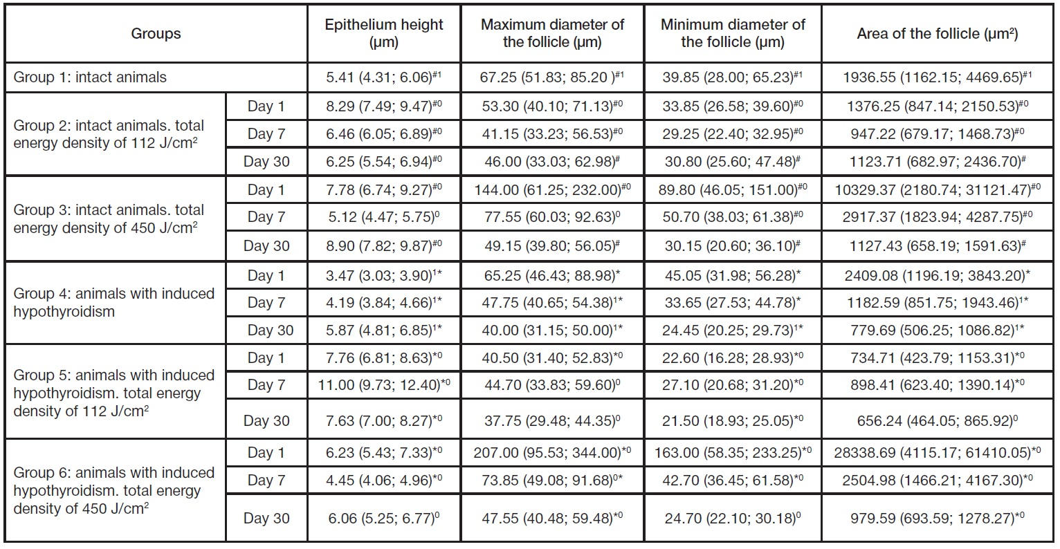
Table 1. Comparative analysis of the epithelium height, diameter and area of follicles in the experimental groups of animals
Note: 1р < 0.05, intact controls compared with animals with untreated induced hypothyroidism; #р < 0.05, irradiated animals compared with intact controls; *р < 0.05, irradiated animals compared with animals with untreated induced hypothyroidism; 0p < 0.05, comparison between the groups of irradiated animals.
Table 2. Comparative analysis of the nucleus/cell ratio and Braun’s index in the experimental groups of animals
Note: 1р < 0.05, intact controls compared with animals with untreated induced hypothyroidism; #р < 0.05, irradiated animals compared with intact controls; *р < 0.05, irradiated animals compared with animals with untreated induced hypothyroidism; 0р < 0.05, comparison between the groups of irradiated animals.