ORIGINAL RESEARCH

A genetically encoded biosensor roKate for monitoring the redox state of the glutathione pool

About authors

1 Shemyakin-Ovchinnikov Institute of Bioorganic Chemistry, Moscow, Russia

2 The Research Institute for Translational Medicine, Pirogov Russian National Research Medical University, Moscow, Russia

Correspondence should be addressed: Dmitry S. Bilan
Miklouho-Maclay, 16/10, Moscow, 117997; moc.liamg@nalib.s.d

About paper

Funding: this work was supported by the Russian Foundation for Basic Research (Project mol_a_dk No.16-34-60175).

Author contribution: Shokhina AG was responsible for the experimental part of the study. Belousov VV and Bilan DS supervised the study and prepared this manuscript.
Received: 2018-12-26 Accepted: 2019-03-02 Published online: 2019-03-15
|
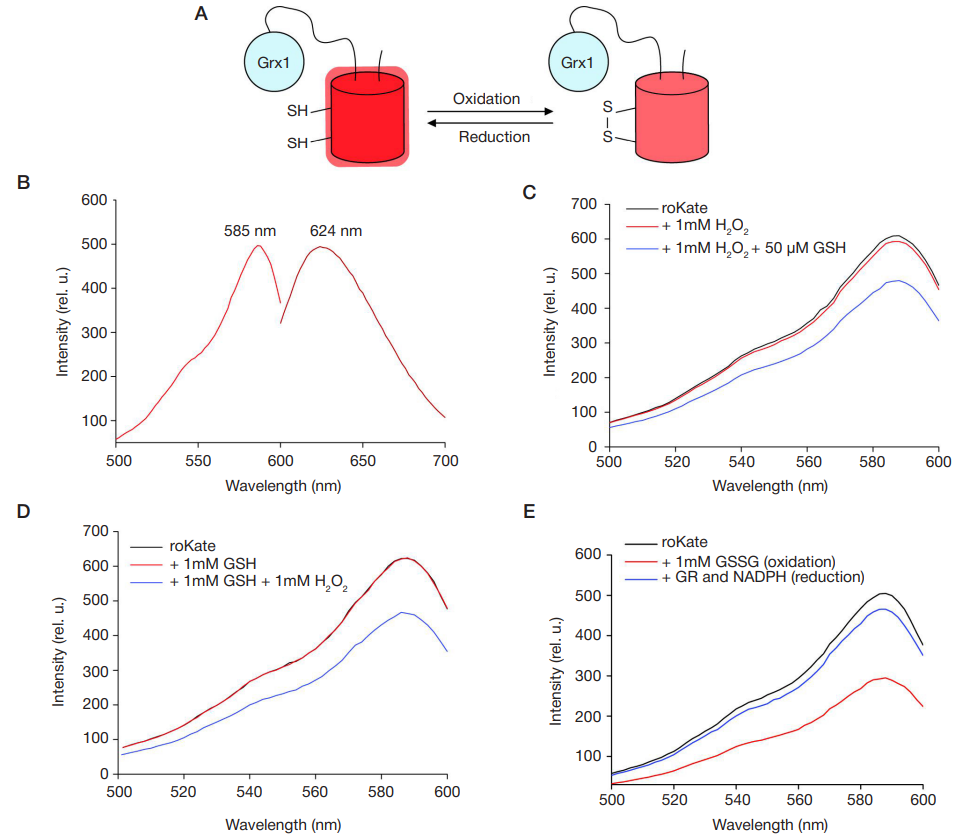
Fig. 1. А. A schematic representation of roKate’s functioning. Two closely positioned cysteine amino acid residues were incorporated into the protein’s structure. When oxidized, the residues form a disulfide bond in the presence of human glutaredoxin 1 (Grx1). Oxidation is accompanied by a drop in fluorescence intensity. B. The fluorescence emission and excitation spectra of the biosensor. C. Changes in the fluorescence excitation spectra in response to the presence of hydrogen peroxide and the subsequent addition of GSH. D. Changes in the fluorescence excitation spectra in response to the presence of GSH and the subsequent addition of hydrogen peroxide. E. Changes in the fluorescence excitation spectra in the isolated protein sample accompanying oxidation (the addition of GSSG) and subsequent reduction (enzymatic reaction of glutathione reduction in the presence of GR and NADPH)
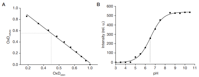
Fig. 2. А Relationship between roKate and glutathione oxidation degrees. B. Relationship between roKate’s fluorescence intensity and pH
Fig. 3. The roKate biosensor in HeLa Kyoto cells. А. Fluorescence intensity dynamics of roKate (the black curve), its variant without Grx1 (the blue curve) and its variant without redox-active cysteine residues (the red curve) in the cytoplasm of living HeLa Kyoto cells in response to the addition of hydrogen peroxide to the medium (the moment hydrogen peroxide was added is marked by an arrow). Error bars show the standard deviation from the mean. B. Images of HeLa Kyoto cells expressing the roKate biosensor before and after the addition of hydrogen peroxide (the time on the images corresponds to that on the graph). The images are shown in pseudo colors corresponding to signal intensity. Scale: 40 μm