ORIGINAL RESEARCH

The state of cognitive functions after angioreconstructive operations on the carotid arteries

About authors

Research Center of Neurology, Moscow, Russia

Correspondence should be addressed: Roman B. Medvedev
Volokolamskoye shosse 80, Moscow, 125367; ur.xednay@namor-vedevdem

About paper

Funding: the study was ordered by the Research Center of Neurology (Federal Research Institution).

Author contribution: Tanashyan MM — study design development, manuscript editing; Medvedev RB — literature analysis, study design development, data collection, analysis and interpretation, manuscript authoring; Lagoda OV and Berdnikovich ES — literature analysis, study design development, data collection and interpretation, manuscript editing; Skrylev SI — angiosurgery, manuscript editing; Gemdzhian EG — study concept and design, data analysis, statistical analysis, manuscript compilation and editing; Krotenkova MV — image data analysis and interpretation, manuscript editing.
Received: 2019-08-15 Accepted: 2019-08-29 Published online: 2019-09-17
|
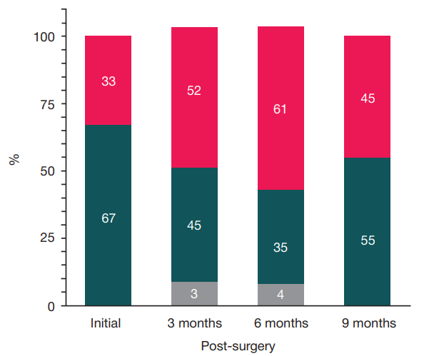
Fig. 1. Dynamics of the proportion of patients (Y axis, %) with different MMSE scores: no cognitive impairment (28–30 score) — upper fields, mild impairment (25–27 score) — middle fields, severe impairment (< 25 score) — two lower fields
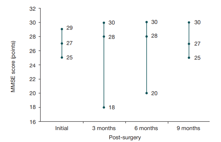
Fig. 2. Dynamics of MMSE scores describing cognitive functions (pre- and post-surgery at different time points); mean values (with minimum and maximum) are presented
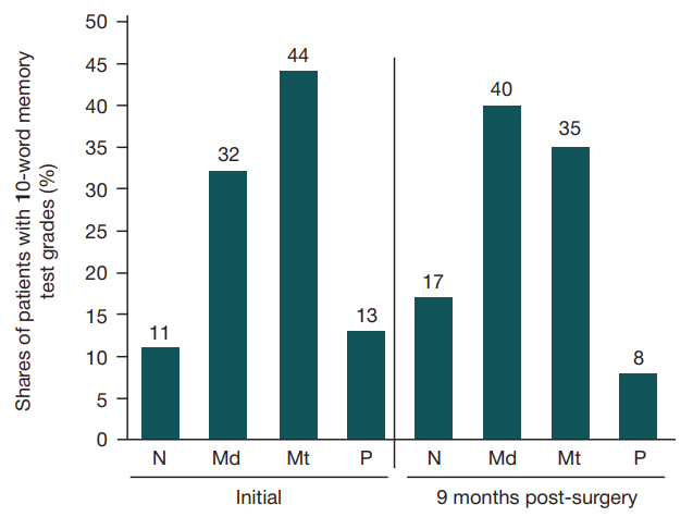
Fig. 3. Operative memory scores (10-word test) compared, pre-surgery (initial) and post-surgery (by the end of the observation period); N — norm (10 words); Md — mild impairments (8–9); Mt — moderate (6–7); S — severe (< 6). By the end of the observation period (9 months) all patients generally exhibited improvements of their operative memory
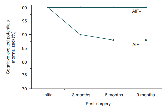
Fig. 4. CEP dynamics in patients with intraoperative acute ischemia foci (AIF+) and in patients without intraoperative acute ischemia foci (AIF–): during the observation period, the CEP level in the AIF+ group (n = 30) did not change, but in the AIF– group (n = 60) it decreased by 15% (p = 0.05). Normalized (divided by the initial levels) main CEP values (%) are given
Fig. 5. Possible changes in the brain matter and post-surgery cognitive functions deterioration vector
Table. Patient CF assessment scores (based on screening tests)*
Note: * — test results are presented as mean values with standard errors of mean (test result histograms are close to normal distribution).