ORIGINAL RESEARCH

Resistive index of internal carotid artery and brain networks in patients with chronic cerebral ischemia

About authors

Research Center of Neurology, Moscow, Russia

Correspondence should be addressed: Vitaly Fedorovich Fokin
Volokolamskoye shosse 80, Moscow, 125367; ur.liam@fvf

About paper

Author contribution: Fokin VF — study concept, manuscript writing; Ponomareva NV — statistical analysis, manuscript writing; Medvedev RB — duplex ultrasonography, hemodynamic data analysis; Konovalov RN — fMRI data acquisition and analysis; Krotenkova MV — fMRI data analysis, study design; Lagoda OV — clinical data analysis; Tanashyan MM — clinical data analysis, study design.

Compliance with ethical standards: the study was approved by the Ethics Committee of the Research Center of Neurology (protocol № 11/14 dated November 19, 2014); the informed consent was submitted by all patients.
Received: 2021-10-27 Accepted: 2021-11-15 Published online: 2021-11-27
|
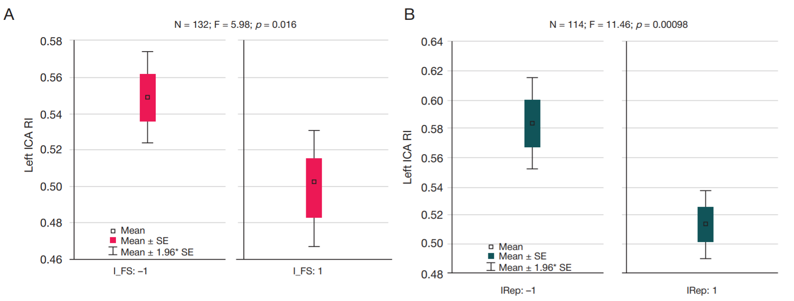
Fig. 1. Left ICA RI in patients with low and high cognitive test scores. А. Left ICA RI in patients with low (–1) and high (1) verbal fluency. B. Left ICA RI in patients with low (–1) and high (1) delayed recall score. I_FS — verbal fluency; IRep — delayed recall; N — number of examined patients; F — F-test; p — significance level; SE — standard error
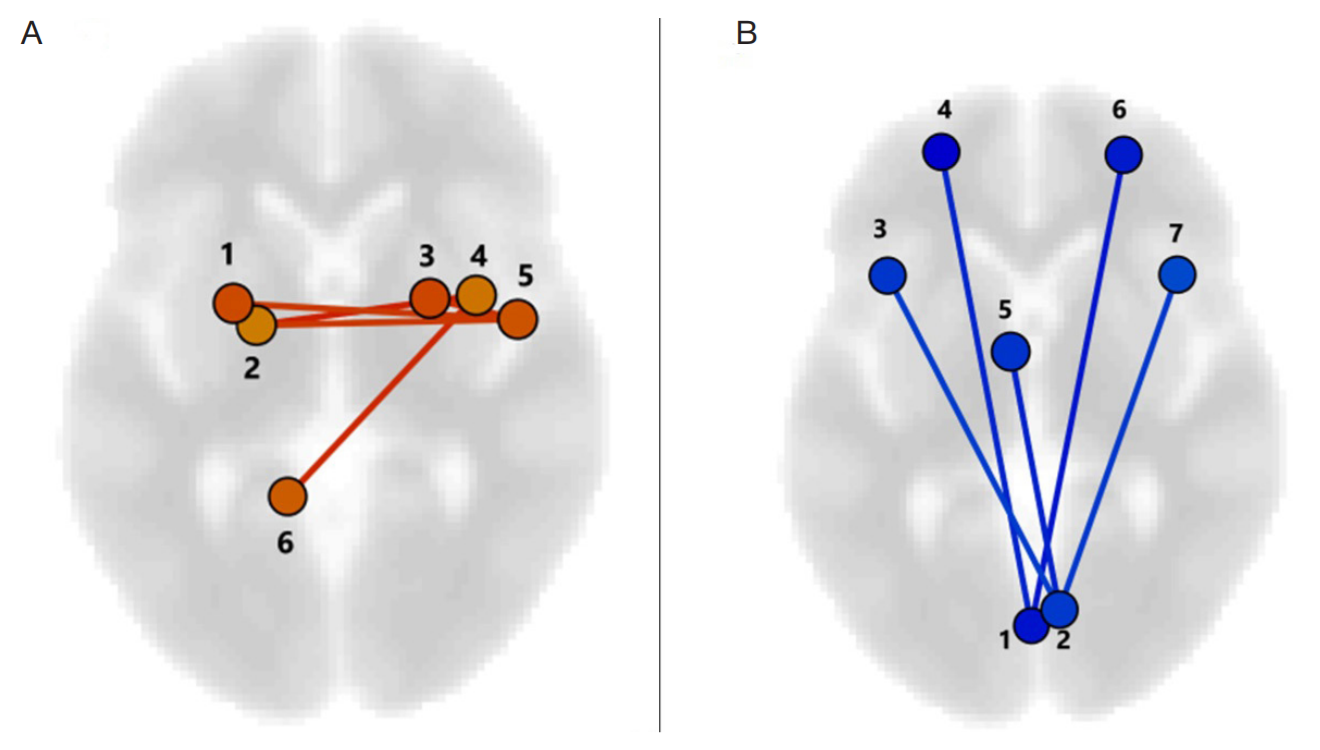
Fig. 2. Differences in connectivity between patients with low (А) and high (B) left ICA RI. А. Connectivity significantly prevailing in patients with low left ICA RI values compared to patients with high RI ( r, l – right and left; 1, 3 – Putamen l, r; 2. – Pallidum; 4 – Insular Cortex (IC); 5 – Planum Polare (PP); 6 – Cerebellum). B. Connectivity significantly prevailing in patients with high left ICA RI values compared to patients with low RI (1 – NetWorks Cerebellar PosteRIor (NW Cereb Post); 2 – SupracalcaRIne Cortex (SCC); 4, 6 – Frontal Pole (FP) l, r; 3, 7 – Frontal Operculum (FO); 5 – Supplementary Motor Cortex (SMC))
Table 1. Correlation between right and left ICA RI values and cognitive functions
Note: n — number of examined patients; p — significance level, females; blank cells correspond to no significant correlation.
Table 2. Significant differences in connectivity between patients with low and high left ICA RI values. А. Positive differences. B. Negative differences
Note: Т — Student’s t-test; p (unadjusted) — significance level unadjusted for multiple comparisons; p (FDR) — significance level adjusted for multiple comparisons; FDR — false discovery rate; other keys are provided in the notes to Fig. 2