ORIGINAL RESEARCH

Medium-term outcomes of extraarticular corrective osteotomy for slipped capital femoral epiphysis

About authors

1 Pirogov Russian National Research Medical University, Moscow, Russia

2 Moscow Regional Clinical Hospital for Trauma and Orthopedics, Moscow, Russia

Correspondence should be addressed: Alexandr V. Grigoriev
Poperechny prosek 3/5, kab. 23, Moscow, Russia; ur.liam@veirogirgva

About paper

Author contribution: all authors contributed equally to the study and the manuscript, all read and approved the final version of the manuscript.

Compliance with ethical standards: the study was approved by the Ethics Committee of Pirogov Russian National Research Medical University and complied with the principles of the Declaration of Helsinki. Informed consent was obtained from the patients’ parents.
Received: 2022-01-10 Accepted: 2022-01-24 Published online: 2022-01-31
|
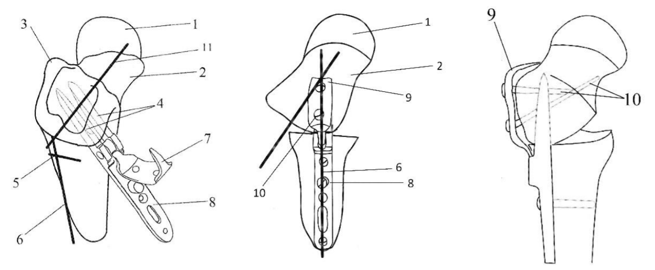
Fig. 1. The schematic representation of the proposed osteotomy technique. 1 — femoral epiphysis; 2 — femoral metaphysis; 3 — the greater trochanter; 4 — jaws of the Trotsenko–Nuzhdin plate; 5 — osteotomy site; 6 — the longitudinal axis of the femoral diaphysis; 7 — the locking part of the plate; 8 — the diaphyseal part of the plate; 9 — the part of the plate anchored to the greater trochanter; 10 — screws fixing the Trotsenko–Nuzhdin plate; 11 — the longitudinal axis of the femur
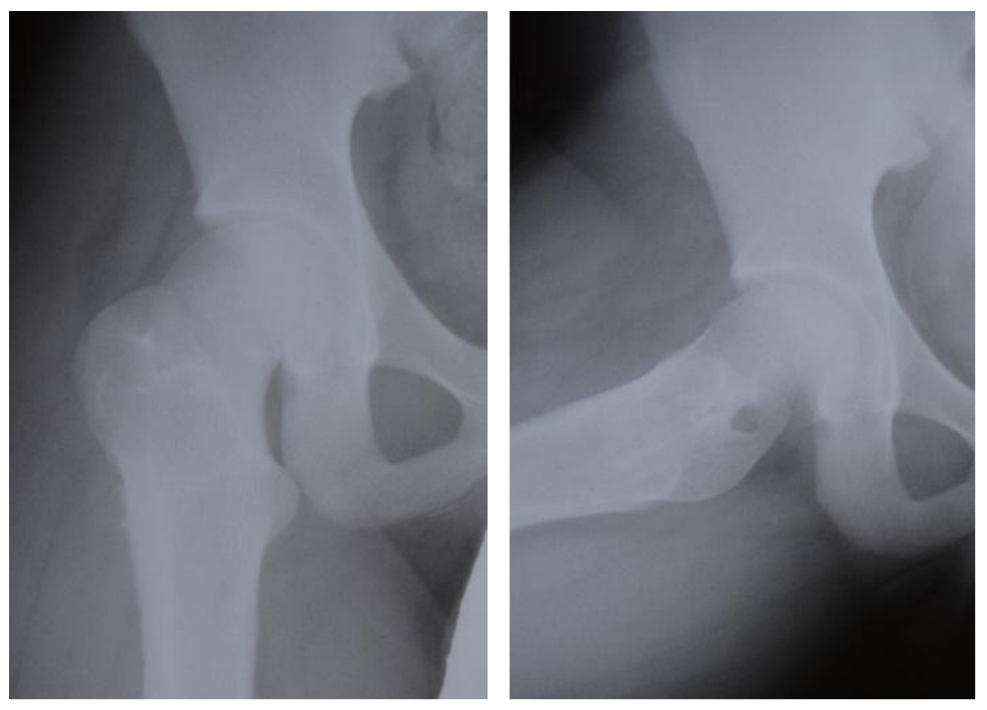
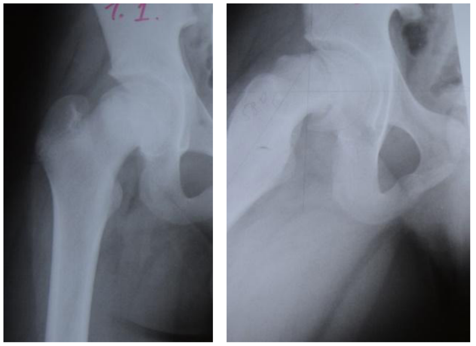
Fig. 2. The anteroposterior and frog-leg lateral radiographs of the right hip (patient M., 12 years). Stage III chronic SCFE
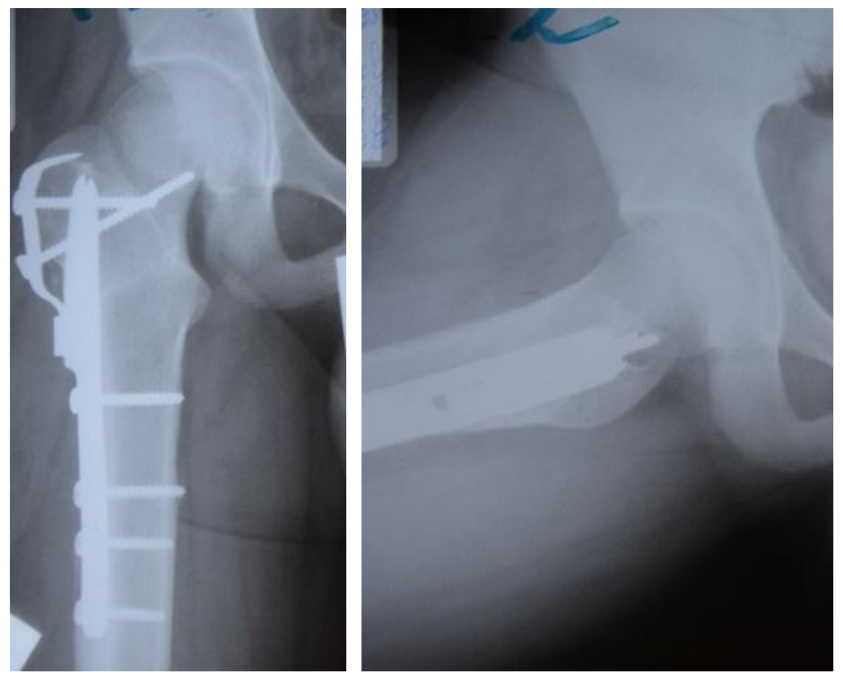
Fig. 3. The anteroposterior and frog-leg lateral radiographs of the right hip (patient M., 12 years) after an intertrochanteric osteotomy of the hip with plate fixation
Fig. 4. The anteroposterior and frog-leg lateral radiographs of the right hip (patient M., 12 years) 12 months after the osteotomy (after bone consolidation and plate removal)
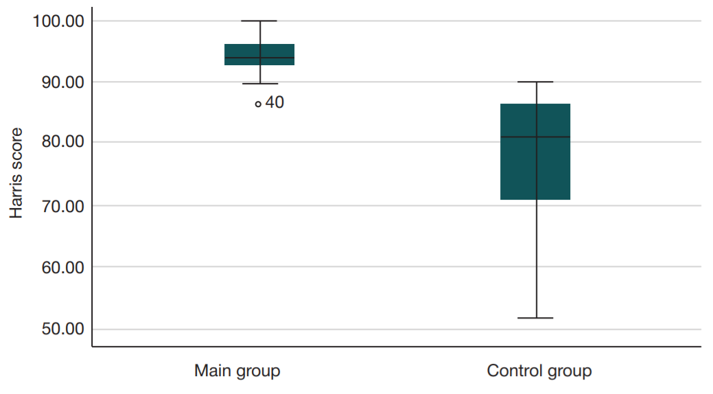
Fig. 5. Harris scores 4.7 years after surgery in the main and control groups
Table. Dynamics of leg length discrepancy 4.7 years after surgery