ORIGINAL RESEARCH

Hemoperfusion and functional state of the macula after simultaneous pancreas and kidney transplantation

Vorobyeva IV1,2, Bulava EV1, Moshetova LK1, Pinchuk AV3,4,5
About authors

1 Russian Medical Academy of Continuous Professional Education, Moscow, Russia

2 Peoples’ Friendship University of Russia, Moscow, Russia

3 Sklifosovskiy Research Institute for Emergency Medical Aid, Moscow, Russia

4 Evdokimov Moscow State University of Medicine and Dentistry, Moscow, Russia

5 Research Institute of Healthcare Organization and Medical Management, Moscow, Russia

Correspondence should be addressed: Irina V. Vorobyeva
Botkinsky pr-d, 2, korp. 19, Moscow, 125284 Russia; ur.liam@0002tnecod.aniri

About paper

Acknowledgements: the authors acknowledge Prof. G.Sh. Arzhimatova of the Botkin Hospital (Moscow) for helpful discussions.

Author contribution: Vorobyeva IV — literature analysis, planning and coordination of the study, data analysis and interpretation; Bulava EV — literature analysis, data collection, analysis and interpretation, preparation of the manuscript; Moshetova LK — study planning and supervision, data analysis and interpretation; Pinchuk AV — study planning and supervision.

Compliance with ethical standards: the study was approved by Ethical Committee at the Russian Medical Academy of Continuous Professional Education (Protocol № 1 of January 18, 2021); the written informed consent for the study was provided by all participants.
Received: 2022-02-22 Accepted: 2022-03-10 Published online: 2022-04-05
|
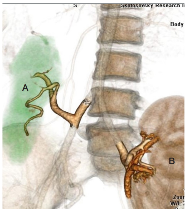
Fig. 1. Computed tomography image with intravenous contrast, 3D reconstruction, showing typical positions of the pancreatic (A) and kidney (B) transplants in the recipient
Fig. 2. Perfusion density measured in four vascular layers of the macular region (color-coded OCT angiography, RS-3000 Advance 2 system, NIDEK, Japan). (А) Superficial capillary plexus of the retina. (B) Deep capillary plexus of the retina. (C) Choriocapillaries. (D) Chorid
Fig. 3. Analysis of functional indicators for the macular region. BCVA — best corrected visual acuity. Group А — patients after simultaneous kidney and pancreas transplantation. Group B — patients awaiting simultaneous kidney and pancreas transplantation. Group C — healthy patients
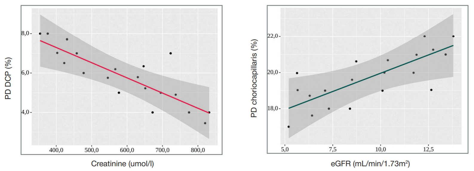
Fig. 4. Regression plots showing correlations of DCP and choriocapillaris perfusion densities with creatinine levels and eGFR, respectively. PD DCP — perfusion density of the deep capillary pexus. PD — perfusion density. EGFR — estimated glomerular filtration rate
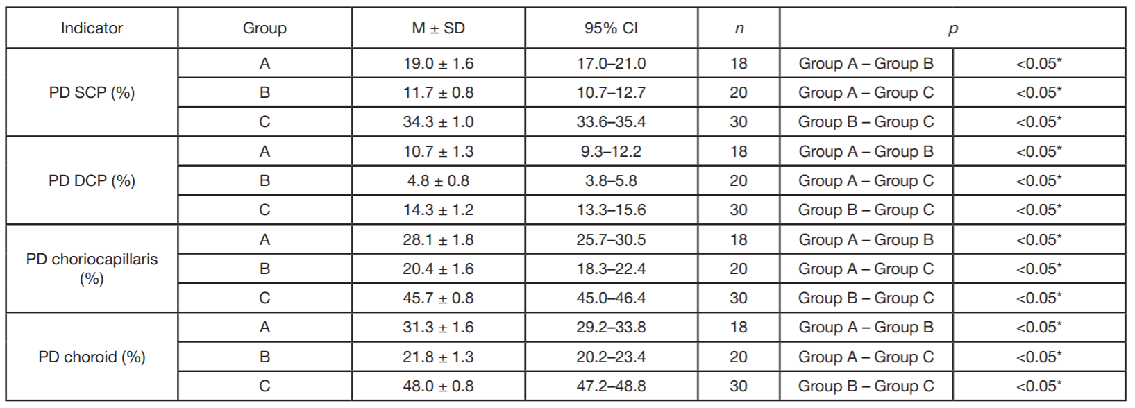
Table. Comparative analysis of perfusion densities in four vascular layers of the macular region
Note: PD SCP — perfusion density of the superficial capillary plexus of the retina; PD DCP — perfusion density of the deep capillary plexus of the retina; PD choriocapillaris — perfusion density of the choriocapillaris; PD choroid — perfusion density of the choroid; Group A — patients after the simultaneous pancreas and kidney transplantation (SPK); Group B — patients on PH/waiting list for the simultaneous pancreas and kidney transplantation (SPK); Group C — healthy individuals.