ORIGINAL RESEARCH

Morphological peculiarities of regeneration of oral mucosa associated with use of polymeric piezoelectric membranes

Koniaeva AD1, Varakuta EYu1, Leiman AE1, Rafiev DO1, Bolbasov EN2, Stankevich KS3
About authors

1 Siberian State Medical University, Tomsk, Russia

2 National Research Tomsk Polytechnic University, Tomsk, Russia

3 Montana State University, Bozeman, MT, USA

Correspondence should be addressed: Anastasiia D. Koniaeva
Moskovsky Trakt, 2, Tomsk, 634034, Russia; moc.liamg@59aynokaysa

About paper

Funding: the study was supported by the Russian Foundation for Basic Research under research project №23-25-00346.

Author contribution: Koniaeva AD, Varakuta EYu, Bolbasov EN, Stankevich KS — study concept and design; Koniaeva AD, Leiman AE — collection and processing of the material; Koniaeva AD, Varakuta EYu, Rafiev DO — text authoring; Koniaeva AD, Varakuta EYu, Rafiev DO, Bolbasov EN, and Stankevich KS — text editing.

Compliance with ethical standards: the study was approved by the Ethics Committee of the Siberian State Medical University (Minutes № 7693/1 of August 26, 2019). All manipulations with the animals were done as prescribed by the Directive 2010/63/EU of the European Parliament of September 22, 2010 "On the protection of animals used for scientific purposes", and the Declaration of Helsinki.
Received: 2023-05-18 Accepted: 2023-06-02 Published online: 2023-06-20
|
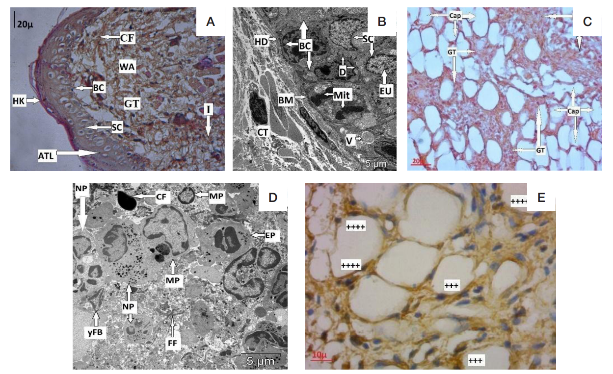
Fig. 1. Rat's buccal mucosa after infliction of the experimental wound defect, 3rd day of the study. A. Newly formed thin epithelial layer in the area of the wound defect and underlying granulation tissue. Pathological changes (acantholysis). Experimental group 2, 3rd day of the study (staining: hematoxylin, eosin; magnification: 400). B. Basal layer cells with signs of intense proliferation and mitotic figures. Experimental group 2, 3rd day of the study (TEM; magnification: 5000). C. New vessels in granulation tissue. Histological picture seen in both experimental groups. Experimental group 2, 3rd day of the study (staining: hematoxylin, eosin; magnification: 400). D. Cellular infiltration in the area of the wound defect. Histological picture seen in both experimental groups. Experimental group 1, 3rd day of the study (TEM; magnification: 5000). E. Expression of VEGF in the endotheliocytes of granulation tissue; nuclei stained with hematoxylin. Experimental group 2, 3rd day of the study (magnification: 900)
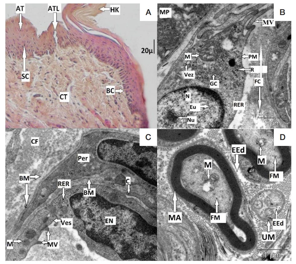
Fig. 2. СRat's buccal mucosa after infliction of the experimental wound defect, 7th day of the study. A. Pathological changes in the epithelium, wound defect in the process of regeneration: unevenly thick epithelial layer, thick spinous layer, acantholysis. Experimental group 1, 7th day of the study (staining: hematoxylin, eosin; magnification: 400). B. Differentiated fibroblast surrounded by connective tissue fibers, wound defect area. Experimental group 2, 7th day of the study (TEM; magnification: 20000). C. Interdigitation between an endothelial cell and a capillary pericyte, wound defect site. Experimental group 2 (TEM; magnification: 20000). D. Peripheral nerve with myelinated and unmyelinated fibers, showing signs of perineural and endoneural edema. Experimental group 2, 7th day of the study (TEM; magnification: 20000).
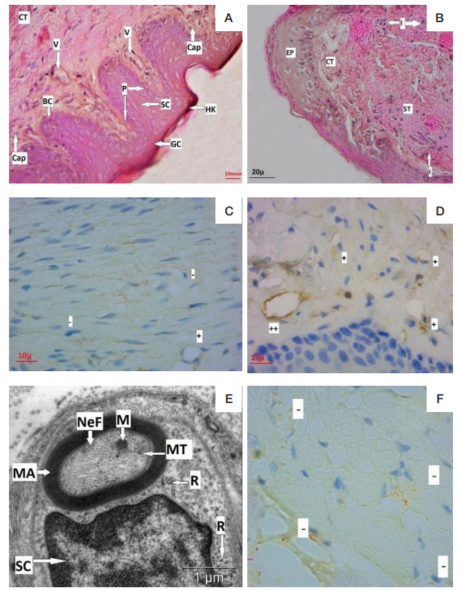
Fig. 3. Rat's buccal mucosa after infliction of the experimental wound defect, 12th day of the study. A. Restored mucosa. Experimental group 2, 12th day of the study (staining: hematoxylin, eosin; magnification: 400). B. Cicatricial tissue, wound defect site. Experimental group 1, 12th day of the study (staining: hematoxylin, eosin; magnification: 400). D, C. Expression of VEGF in the endotheliocytes of granulation tissue; nuclei stained with hematoxylin. C. Experimental group 1. D. Experimental group 2, 12th day of the study (magnification: 900). E. Peripheral nerve with myelin fibers showing no signs of perineural and endoneural edema, wound defect area. Experimental group 2 (magnification: 20000). F. Absence of S-100 expression in the mucosa's lamina propria, nuclei staining with hematoxylin. Histological picture seen in both experimental groups. Experimental group 1, 12th day of the study (magnification: 900). WA — wound defect area; CT — loose fibrous connective tissue; GT — granulation tissue; ST — dense fibrous connective tissue; I — infiltration; V — venule; Cap — capillary; EP — epithelium; BM — basement membrane; BC — basal cells; SC — acanthocytes; GС — surface layer cells; HK — stratum corneum; ATL — acantholysis; AT — acanthosis; P — papillae; D— desmosomes; HD — hemidesmosomes; Va — expansion of intercellular space; PM — plasma membrane; EU — euchromatin; N — core; Nu — nucleolus; RER — granular endoplasmic reticulum; R — polysomes; M — mitochondrion; GC — Golgi complex; MV — outgrowths of plasmolemma; Vez — vesicles; C — intercellular contact; Mit — mitosis; yFB — young fibroblast; MP — macrophage; NP — neutrophil; EP — eosinophil; FC — connective tissue fibers; EN — endotheliocyte; Per — pericyte; FF — fragments of connective tissue fibers; CF — destructively altered cells; CF— collagen fibers; PN — peripheral nerve; MA — myelinated nerve fiber; UM — unmyelinated nerve fiber; PEd — perineural space edema; EEd — edema of the endoneural space; FM — neurofilaments and microtubules; ++++ — very strong staining; +++ —strong staining; ++ — moderate staining; + — weak staining; ‒ — no staining
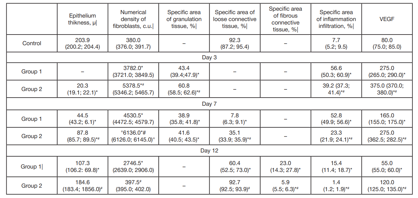
Table. Morphological indicators of changes in the oral mucosa during regeneration of a wound defect, М(Q1:Q3)
Note: * — significant differences compared to the control group (p < 0,05); # — significant differences compared to group 1 (p < 0,05).