Copyright: © 2024 by the authors. Licensee: Pirogov University.
This article is an open access article distributed under the terms and conditions of the Creative Commons Attribution license (CC BY).

ORIGINAL RESEARCH

Comparative analysis of methods for calculation of toric intraocular lenses in patients after penetrating keratoplasty

Sinitsyn MV1,2, Voskresenskaya AA1,2, Pozdeyeva NA1,2
About authors

1 Cheboksary branch of the Fyodorov Eye Microsurgery Federal State Institution, Cheboksary, Russia

2 Postgraduate Doctors’ Training Institute of the Ministry of Public Health of Chuvashia, Cheboksary, Russia

Correspondence should be addressed: Maxim V. Sinitsyn
pr. Traktorostroitelej, 10, 428028, Cheboksary, Russia; ur.liam@nicinisktnm

About paper

Author contribution: Sinitsyn MV — study concept and design, data acquisition, data analysis and processing, statistical analysis, manuscript writing; Voskresenskaya AA, Pozdeyeva NA — editing, approval of the final version of the article.
Received: 2024-11-11 Accepted: 2024-12-01 Published online: 2024-12-22
|
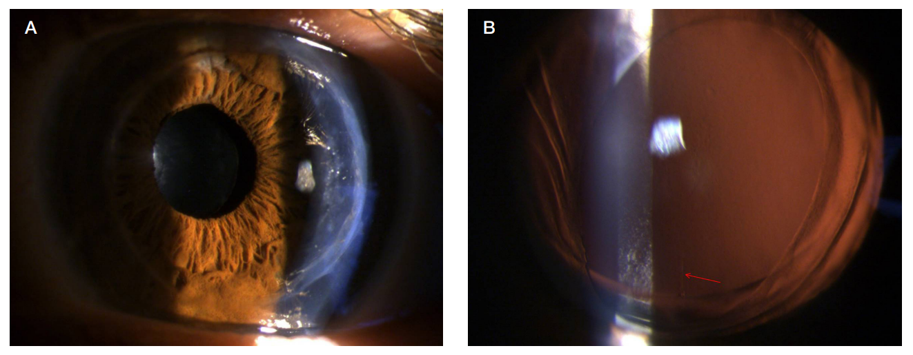
Fig. 1. View of the eye of patient N., 56 years, during slit lamp examination on day 1 after phacoemulsification with the toric intraocular lens implantation. А. A transparent corneal graft can be seen, along with the optic part of the toric intraocular lens in the projection of the pupil. B. View of the eye against the background of drug-induced mydriasis, a vertical mark of the intraocular lens toric axis (marked with a red arrow) located along the corneal graft keratometry strong meridian can be seen
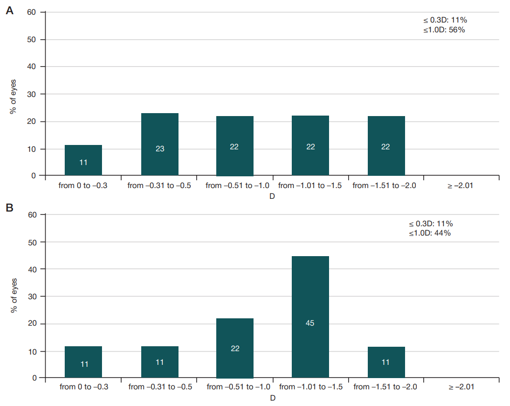
Fig. 2. Diagrams of the distribution of pre-operative values of corneal astigmatism on the posterior corneal graft surface based on the corneal OCT data (CASIA 2) (А), Scheimpflug keratotopography data (Pentacam) (B)
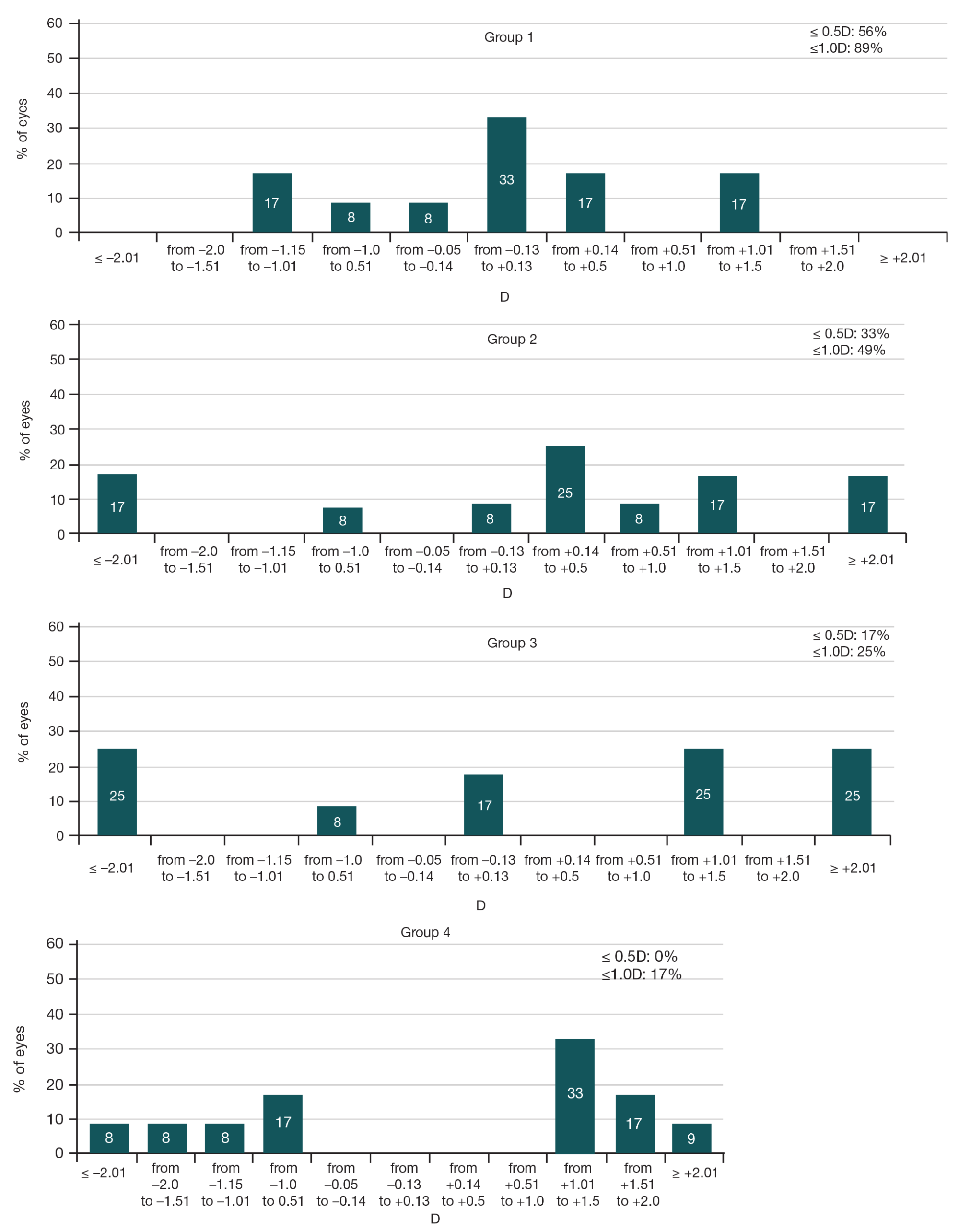
Fig. 3. Predictability of the spherical refraction component when calculating the tIOL optic power in groups 1–4
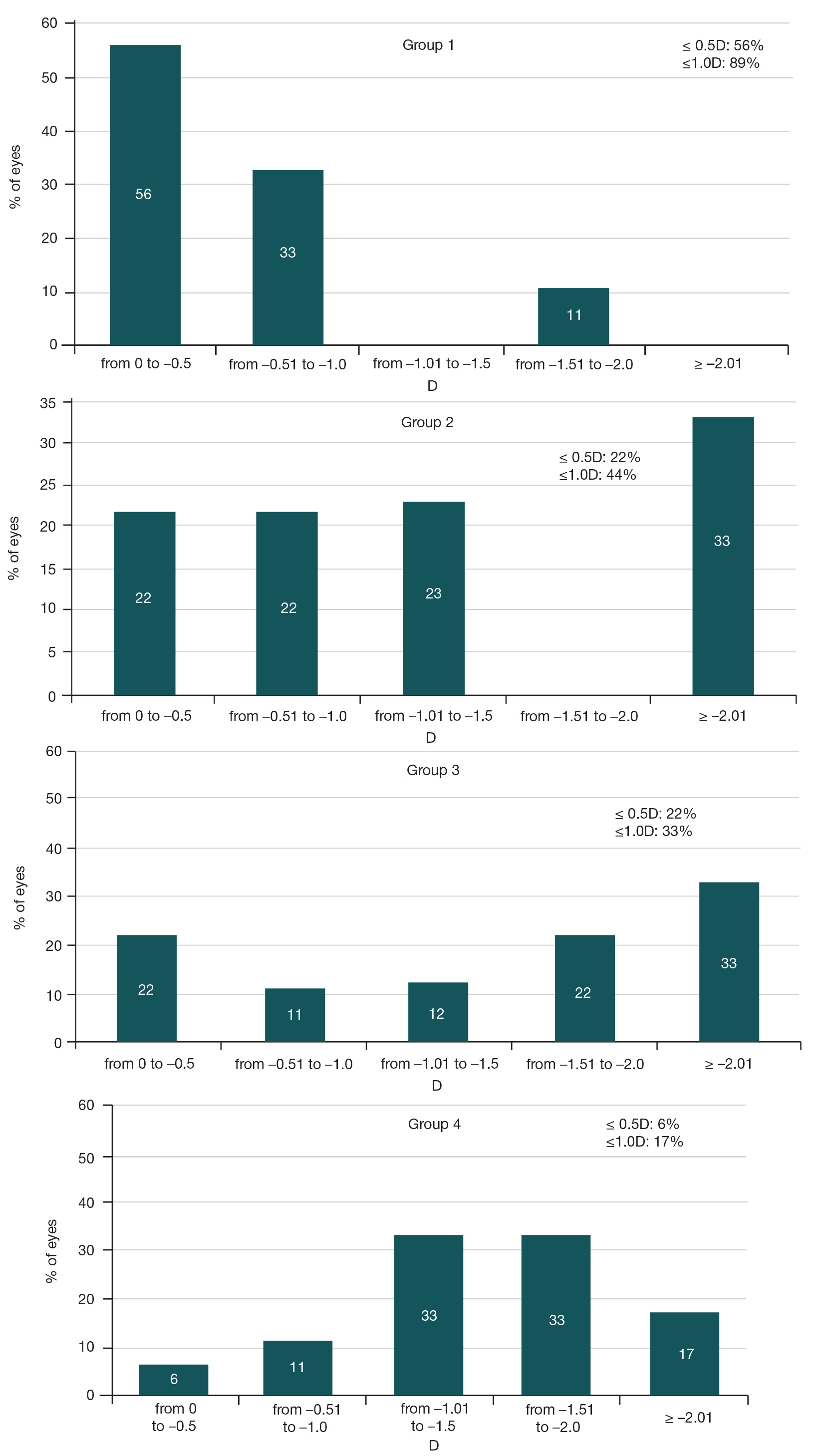
Fig. 4. Predictability of the cylindrical refraction component when calculating the tIOL optic power in groups 1–4
Table 1. Methods for toric intraocular calculation during cataract extraction in patients after penetrating keratoplasty
Table 2. Preoperative values of corneal astigmatism on both corneal graft surfaces measured using different equipment (M ± σ)
Table 3. Preoperative average keratometry values of both corneal graft surfaces measured using different equipment (M ± σ)