Copyright: © 2025 by the authors. Licensee: Pirogov University.
This article is an open access article distributed under the terms and conditions of the Creative Commons Attribution license (CC BY).

ORIGINAL RESEARCH

Morphological, immunohistochemistry and molecular analysis of differentiated high-grade carcinoma

Makhachev DR, Bulanov DV, Shovkhalov MM, Bekmurziev BZ, Geroev IA, Netsvetova AM, Zhusupova AR, Gubich DS, Manovski AM
About authors

Pirogov Russian National Research Medical University (Pirogov University), Moscow, Russia

Correspondence should be addressed: Dalgat R. Makhachev
Akademika Volgina, 37, Moscow, 117437, Russia; ur.liam@2002taglad

About paper

Author contribution: FMakhachev DR, Bulanov DV, Shovkhalov MM, Bekmurziev BZ, Geroev IA — data analysis and interpretation, manuscript writing, editing; Netsvetova AM, Zhusupova AR — manuscript writing, editing; Gubich DS, Manovski AM — clinical data acquisition, editing.
Received: 2025-09-02 Accepted: 2025-09-15 Published online: 2025-09-24
|
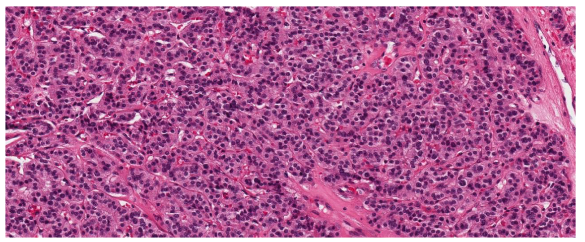
Fig. 1. Poorly differentiated thyroid carcinoma (PDTC) based on the Turin Proposal criteria. Solid/insular growth pattern with thin fibrovascular septa, highly dense small monomorphic cells with high nuclear/cytoplasmic ratio; no nuclear features typical for papillary thyroid carcinoma (H&E, ×200 magnification)
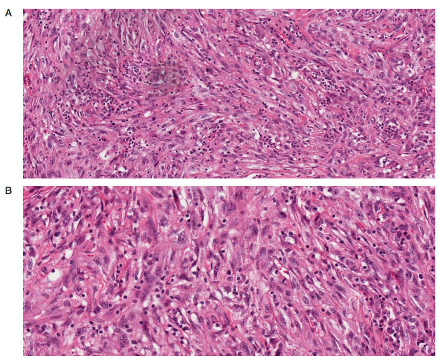
Fig. 2. Spindle cell variant of anaplastic thyroid carcinoma (ATC). A. Dense, multidirectional bundles of spindle cells with the pronounced nuclear pleiomorphism, coarsely granular chromatin (H&E, ×100 magnification). B. High cellularuty, multiple atypical mitoses, sporadic multinucleated tumor cells (H&E, ×400 magnification)
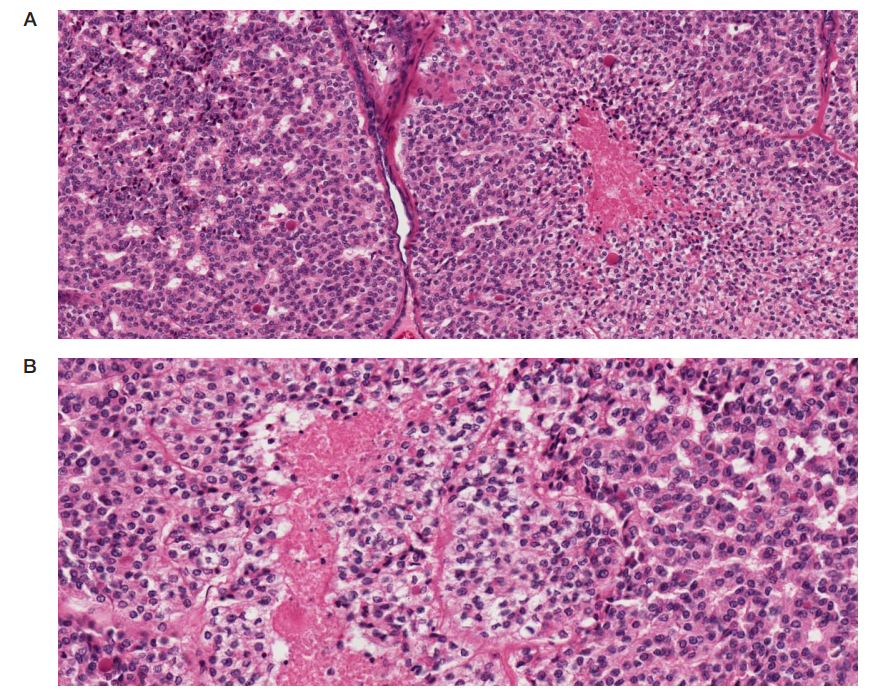
Fig. 3. Differentiated high-grade thyroid carcinoma (DHGTC). A. Solid/trabecular and alveolar areas with the zone of comedo/geographical necrosis (H&E, ×100 magnification). B. Pronounced increase in cytological atypia and proliferation; frequent mitoses (≥ 5 per 2 mm2 when counted in the fields with the highest activity) (H&E, ×400 magnification)
Table. Comparison of morphological, immunohistochemical, and molecular features of poorly differentiated (PDTC) and differentiated high-grade tyroid carcinoma (DHGTC)