Copyright: © 2025 by the authors. Licensee: Pirogov University.
This article is an open access article distributed under the terms and conditions of the Creative Commons Attribution license (CC BY).

ORIGINAL RESEARCH

Comparative analysis of metallic endovascular coil frame designs

Chepeleva EV1, Kozyr KV1, Borodin VP1, Khakhalkin VV1, Vladimirov SV1, Makhmudov MA1, Badoian AG1, Baranov AA1, Krestyaninov OV1,2
About authors

1 Meshalkin National Medical Research Center, Novosibirsk, Russia

2 Novosibirsk State Medical University, Novosibirsk, Russia

Correspondence should be addressed: Elena V. Chepeleva
Rechkunovskaya, 15, Novosibirsk, 630055, Russia; ur.niklahsem@avelepehc_e

About paper

Funding: the study was conducted as part of the Russian Science Foundation project No. 25-15-00480.

Author contribution: Chepeleva EV — data analysis, manuscript writing and editing; Kozyr KV, Borodin VP — experimental procedure, data analysis, visualization of findings; Khakhalkin VV — study concept and design, data analysis; Vladimirov SV — experimental procedure, data analysis, visualization of findings; Makhmudov MA, Badoian AG, Baranov AA — technical support, data validation, graphics design; Krestyaninov OV — general research management, coordinating work, editing and approval of the final version of the manuscript.
Received: 2025-09-15 Accepted: 2025-09-30 Published online: 2025-10-23
|
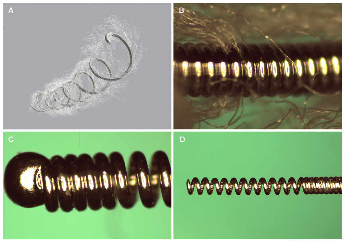
Fig. 1. Nester endovascular coil manufactured by COOK MEDICAL LLC: general view (A); fiber attachment (B); atraumatic tip (C); interlock part (D)
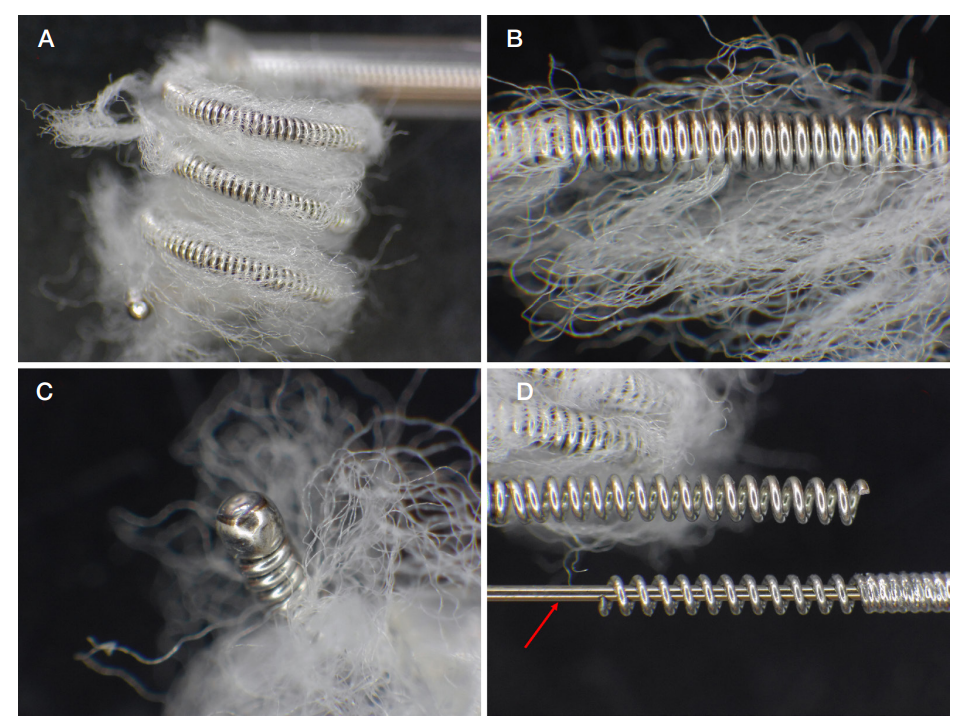
Fig. 2. MReye Flipper endovascular coil manufactured by COOK MEDICAL LLC: general view (A); fiber attachment (B); atraumatic tip (C); interlock parts of the coil and delivery system, delivery system rod (red arrow) (D)
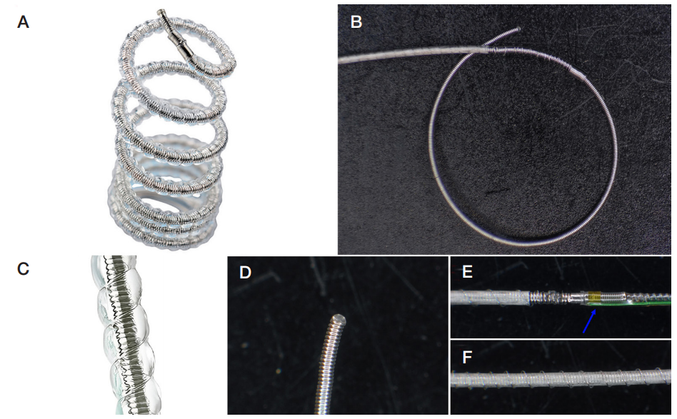
Fig. 3. Terumo AZUR 18 endovascular coil manufactured by MicroVention, Inc.: general view (A); distal part of the coil, in which the margin, where the hydrogel sheath begins, is pointed with the red arrow (B); activated hydrogel sheath (C); atraumatic tip (D); thermoelectric coil detachment system (blue *) (E); hydrogel sheath (F)
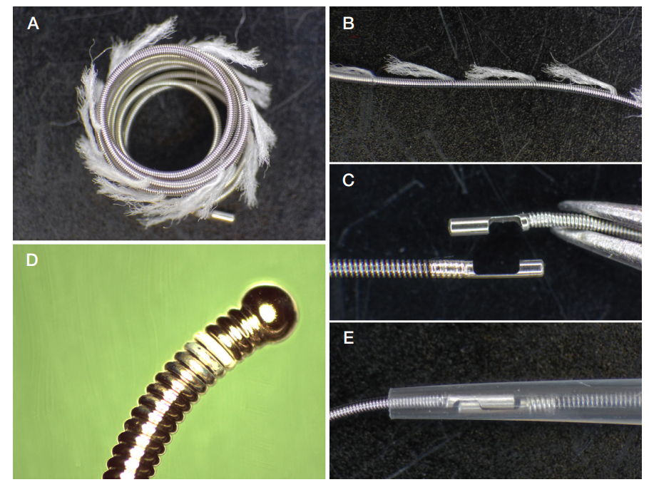
Fig. 4. Interlock Spiral 2D endovascular coil manufactured by Boston Scientific Corporation: general view (A); fiber attachment (B); interlock parts of the coil and delivery system (C); atraumatic tip (D); interlocked coil and delivery system (E)
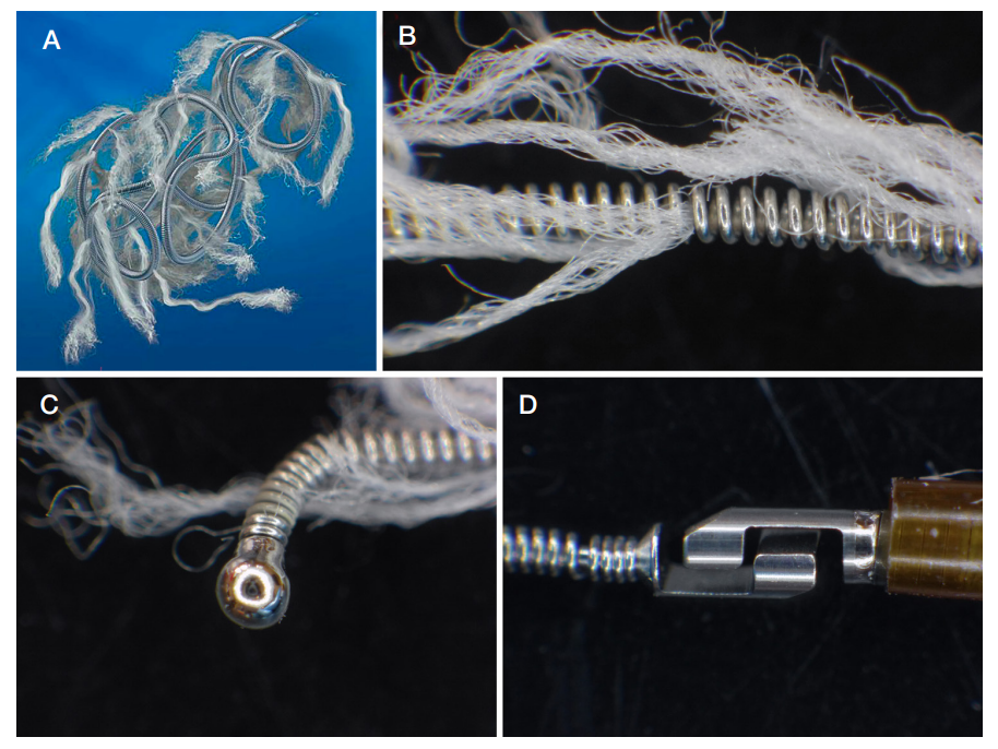
Fig. 5. Interlock Spiral - 35 endovascular coil manufactured by Boston Scientific Corporation: general view (A); fiber attachment (B); atraumatic tip (C); interlock part of the coil (D)
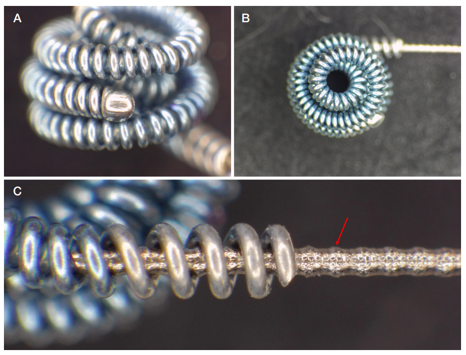
Fig. 6. Nit-Occlud PDA endovascular coil manufactured by PFM Medical GMBH: atraumatic tip (A); general view (B); interlock part, in which the screw notches on the delivery system rod for connection to the coil are pointed with the red arrow (C)
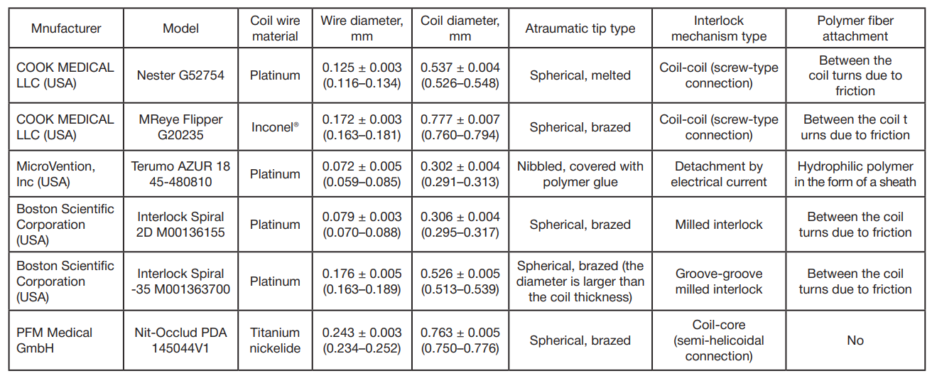
Table. Endovascular coil characteristics. Values are presented as M ± SD, where M is the mean; SD is the standard deviation; the 95% confidence interval for each mean is provided in parentheses.