METHOD

Iodine quantification with computed tomography for the purpose of dose assessment in contrast enhanced radiotherapy

Lipengolts AA1,2,3, Budaeva JA2,3, Blaickner M4, Cherepanov AA1, Menkov MA1, Kulakov VN2, Grigorieva EYu1
About authors

1 N. N. Blokhin Russian Cancer Research Center, Moscow, Russia

2 A. I. Burnazyan Federal Medical and Biophysical Center, Moscow, Russia

3 National Research Nuclear University MEPhI, Moscow, Russia

4 Austrian Institute of Technology, Vienna, Austria

Correspondence should be addressed: Alexey A. Lipengolts
Kashirskoe shosse, d. 24, Moscow, Russia, 115478; ur.liam@stlognepil
Received: 2016-12-01 Accepted: 2016-12-06 Published online: 2017-01-19
|
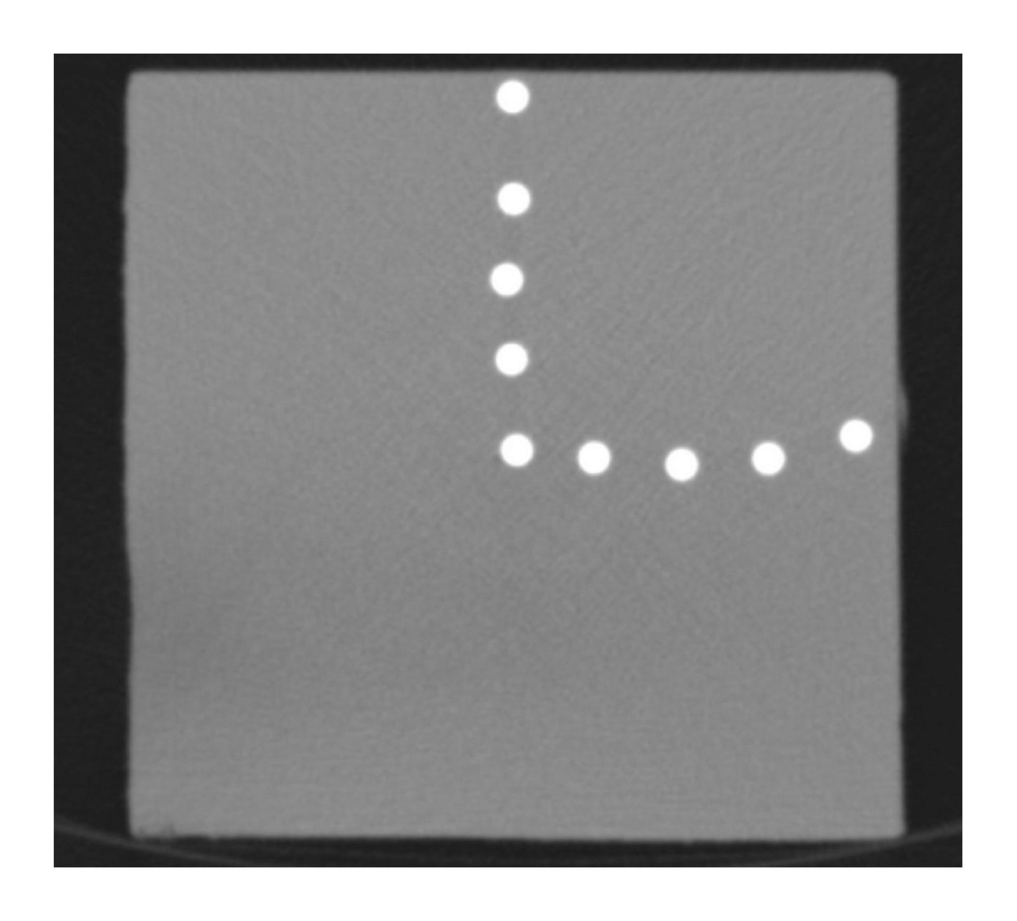
Fig. 1. A CT image of a polyethylene phantom with microtubes in it containing a 35 mg/ml aqueous iodine solution. Operating voltage is 80 kV
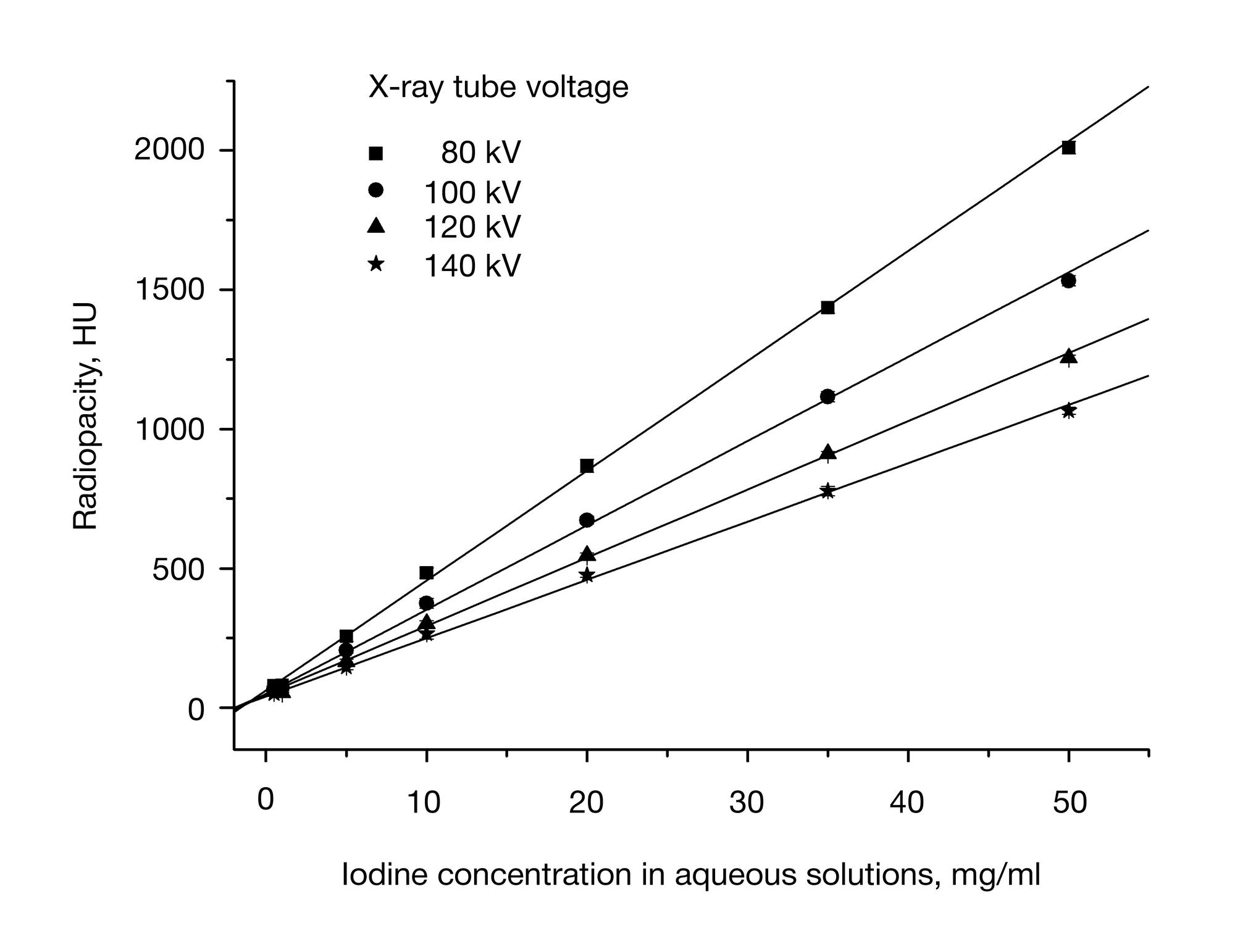
Fig. 2. Calibration curves showing the relationship between radiopacity (expressed in Hounsfield units, HU) and iodine concentrations at X-ray tube voltages of 80, 100, 120 and 140 kV for solutions placed inside the phantom
Iodine concentrations in aqueous solutions measured by a CT scanner and their deviations from actual values at various voltages