CLINICAL CASE

Formation of arteriovenous fistula following surgical resection of vestibular schwannoma

Reutov AA1, Aronov MS2, Kushel YuV3
About authors

1 Clinical Hospital of the Department for Presidential Affairs of the Russian Federation, Moscow

2 Burnasyan Federal Medical Biophysical Center, Moscow, Russia

3 Burdenko National Medical Research Center of Neurosurgery, Moscow

Correspondence should be addressed: Andrey A. Reutov
Marshala Timoshenko 15, Moscow, 121359; ur.oruenretnec@votuer
Received: 2018-01-19 Accepted: 2018-02-20 Published online: 2018-08-15
|
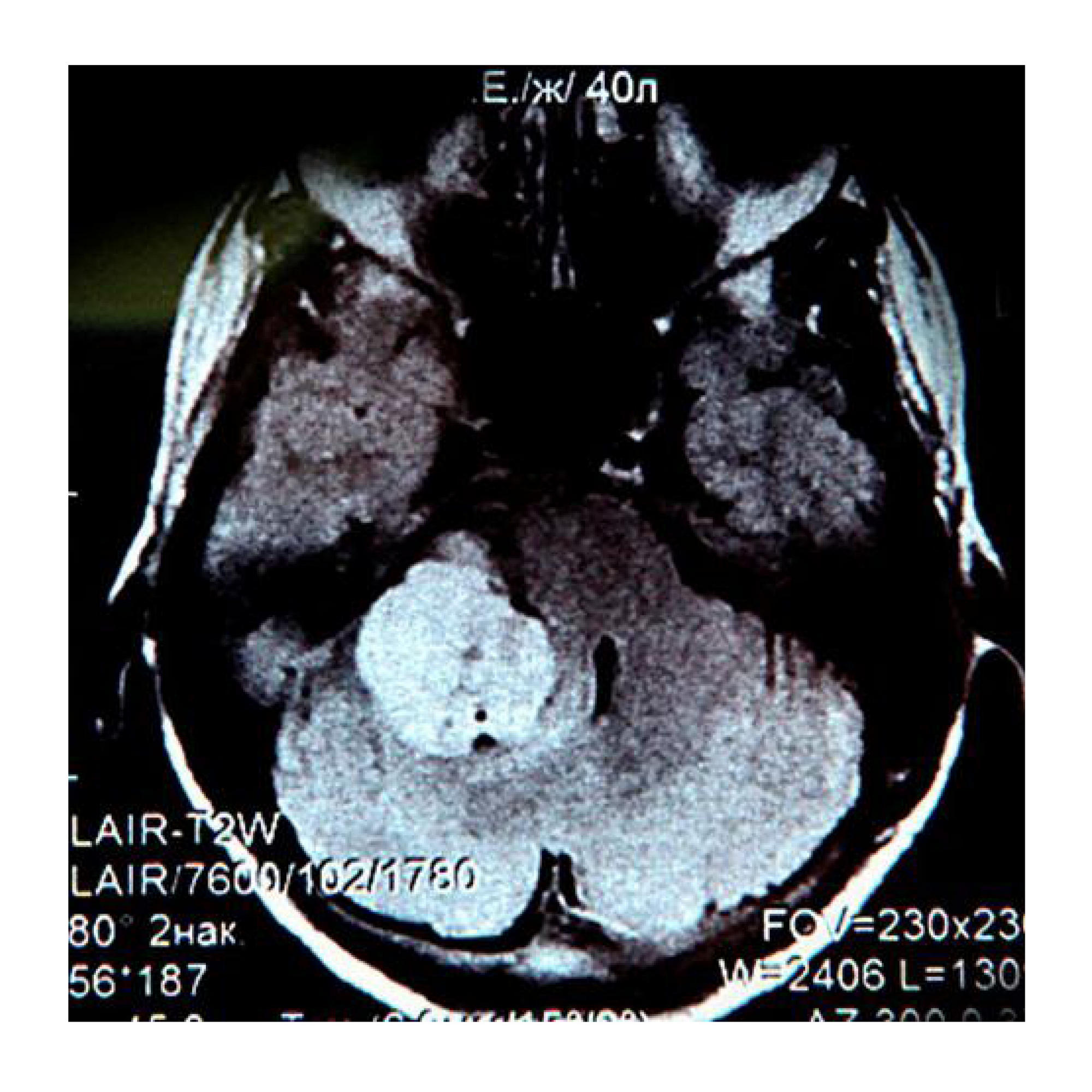
Fig. 1. The T1-weighted contrast-enhanced MR image taken before the surgery shows a typical right-sided vestibular schwannoma
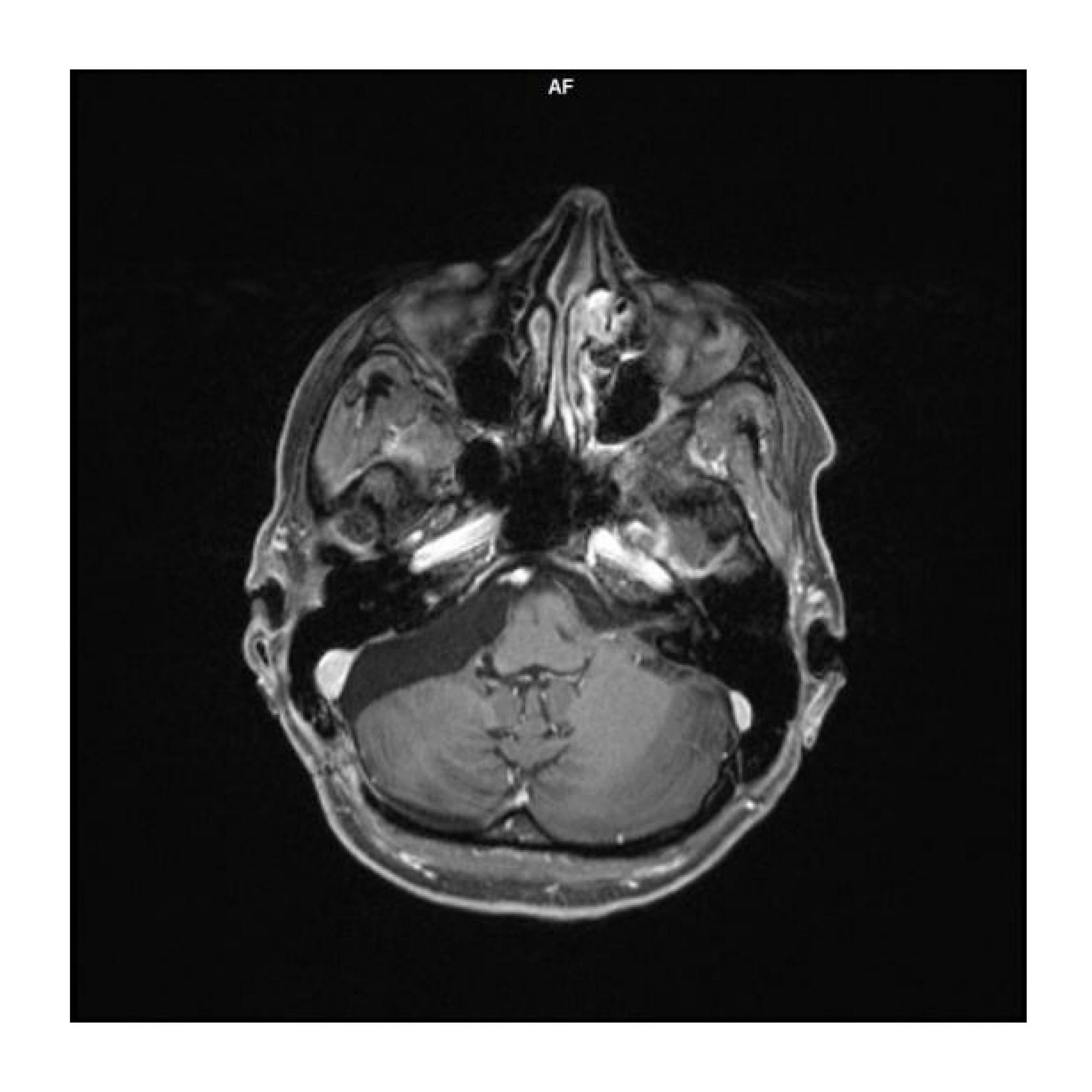
Fig. 2. The T1-weighted contrast-enhanced MR image taken after the surgery shows no tumor remnants or postoperative complications
Fig. 3. Postoperative magnetic resonance angiography images showing no vascular damage
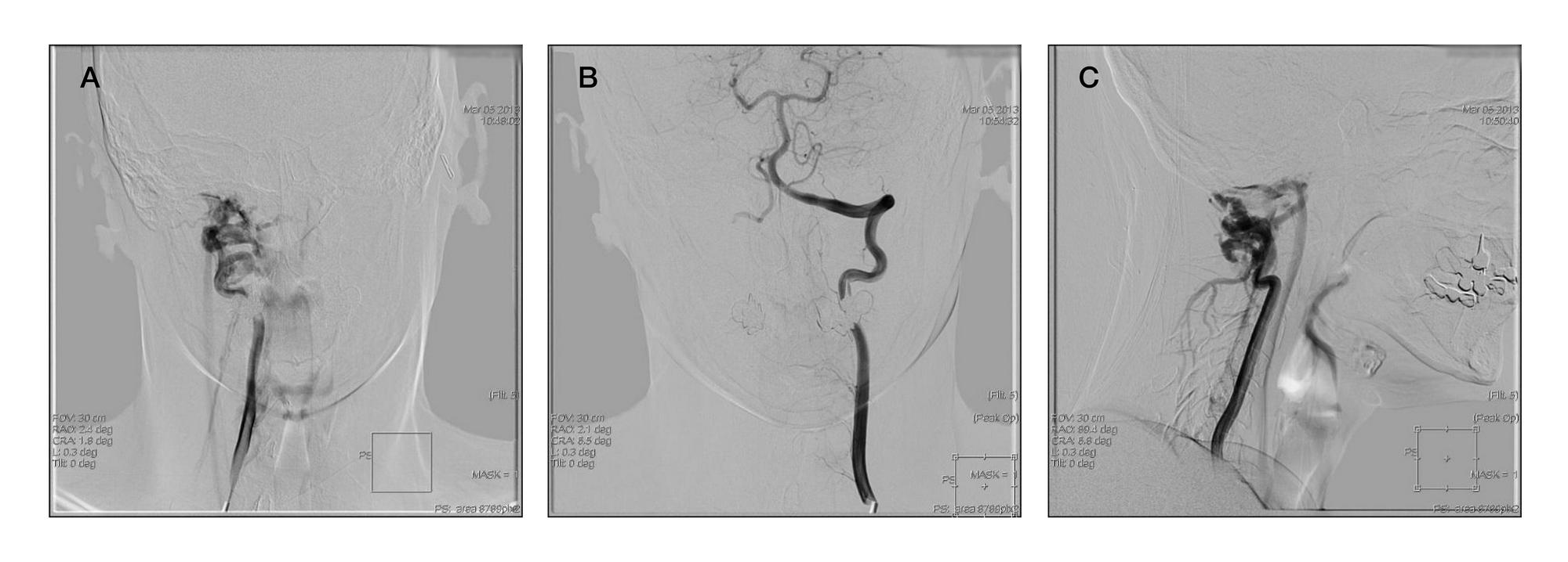
Fig. 4. The initial angiogram of the right vertebral artery (A), left vertebral artery, frontal view (B), and right vertebral artery, lateral view (C)
Fig. 5. The intraoperative angiogram, of the right (A, C) and left (B) vertebral arteries, frontal view. Retrograde opacification of the fistula via the left vertebral artery is observed
Fig. 6. Occlusion of the distal segment of the right vertebral artery via the collateral approach
Fig. 7. Native (A) and subtraction (B, C) angiograms of the left vertebral artery appearing in the frontal (C) and lateral (A, B) views. The vertebrobasilar system is totally contrast-opacified while the fistula is not