ORIGINAL RESEARCH

Activation of CD4+CD39+ Т cells in colorectal canser

Zhulai GА1, Churov AV1, Oleinik EK1, Romanov AA2, Semakova PN1, Oleinik VM1
About authors

1 Institute of Biology, Karelian Research Center of the Russian Academy of Sciences (IB KarRC RAS), Petrozavodsk

2 Republic Oncology Center, Petrozavodsk

Correspondence should be addressed: Galina A. Zhulai
Pushkinskaya 11, Petrozavodsk, Republic of Karelia, 185014; ur.xednay@111-ilaghz

About paper

Funding: the study was part of the State assignment for the Federal Research Center Karelian Research Center of the Russian Academy of Sciences (Project 0221-2017-0043).

Received: 2018-01-25 Accepted: 2018-03-20 Published online: 2018-07-25
|
Fig. 1. Levels of CD4+CD39+ T cells in the peripheral blood of patients with CRC
Fig. 2. Levels of CD39-expressing lymphocytes in the peripheral blood and tumor tissue of patients with CRR
Note: * — represents significant differences at p < 0.05.
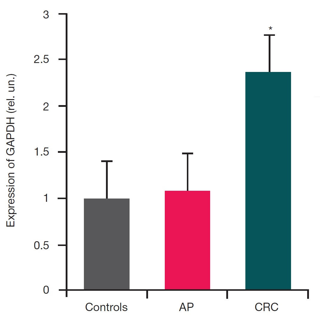
Fig. 3. Changes in the levels of CD39 mRNA in the peripheral blood leukocytes of patients with CRC and AP (relative to GAPDH mRNA)
Note: * — represents significant differences from the controls at p < 0.05; data are presented as М ± SE.
Table 1. Percentage of major lymphocyte subsets in the peripheral blood of healthy donors, patients with CRC and patients with AP from the total number of lymphocytes
Note: * — represents significant differences from the controls, p < 0.05; # — represents significant differences from patients with stages I and II CRC, p < 0.05.
Table 2. Expression of ectonucleotidase C39 in the subset of CD4+ Т cells isolated from patients with CRC presented as percentage from the total number of CD4+ Т cells
Note: * — represents significant differences from the controls; ** — represents significant differences from the controls and patients with stages I and II CRC; # — represents significant differences from the peripheral blood lymphocytes of the same patients with CRC.