ORIGINAL RESEARCH

Nanoparticles guided precise transplantation of varying numbers of mesenchymal stem cells into post–traumatic syrinx in spinal cord injury rat

Zhang C1,2,3,4, Morozova AYu4, Baklaushev VP3, Gubsky IL2, Melnikov PA4, Gabashvily AN6, Wang G1, Li L1, Wu H1, Wang X5, Chekhonin VP2,4
About authors

1 Department of Bone and Soft Tissue Tumors, Tianjin Medical University Cancer Institute and Hospital, National Clinical Research Center for Cancer, Key Laboratory of Cancer Prevention and Therapy, Tianjin’s Clinical Research Center for Cancer, Tianjin, China

2 Department of Medicinal Nanobiotechnology, Pirogov Russian National Research Medical University, Moscow

3 Federal Medical and Biological Agency, Moscow

4 Department of Basic and Applied Neurobiology, Federal Medical Research Center for Psychiatry and Narcology, Moscow

5 Department of Epidemiology and Biostatistics, First Affiliated Hospital, Army Medical University, Chongqing, China

6 Vavilov Institute of General Genetics Russian Academy of Sciences, Moscow

Correspondеnce should be adressed: Chao Zhang
Huanhu Xi Road, Hexi Area, Tianjin, 300060, China; nc.ude.umt@oahcgnahzrd; gro.noitadnuofoa@sdrahcir.ffoeg

About paper

Funding: the present study was sponsored by China Scholarship Council (201406940004), and Russian Scientific Foundation (16–15–10432).
Received: 2018-08-26 Accepted: 2018-09-25 Published online: 2018-12-31
|
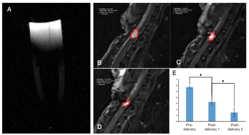
Fig. 1. Nanoparticles transplantation and MRI analysis. A. Nanoparticles showed low signal change in T2–weighted scanning. B–E. The volume change of PTS post nanoparticle transplantation. PTS volume decreased after the first transplantation, and further decreased after the second transplantation (p < 0.05)
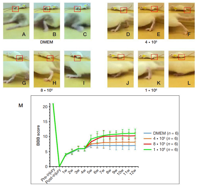
Fig. 2. Motor function recovery evaluated by BBB score. Differences in the BBB scores among the four groups were not statistically significant before cell transplantation. One–week post transplantation, compared with the DMEM group, the differences were found to be statistically significant in cell transplanted groups (M). Compared with the DMEM group, the behavior ability suggested the better outcome in the cell transplanted groups. Among all the groups, 1 • 106 group showed the best outcome (M). Eight–week post transplantation, the rats showed different abilities on weight support (A, D, G, J), plantar placement (B, E, H, K), and plantar stepping (C, F, I, L)
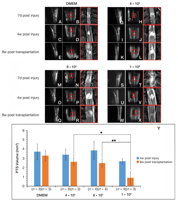
Fig. 3. The Reduction of post-traumatic syrinx. Lateral and horizontal MRI scanning was performed in all groups to evaluate the situation of post-traumatic syrinx preand post- transplantation (A–X). The volume of PTS decreased in the cell transplant groups when evaluated 8 w post transplantation. Among all the groups, 1 • 106 group showed the best improvement (S–Y). The areas in the red box were magnified in the figures with red frame
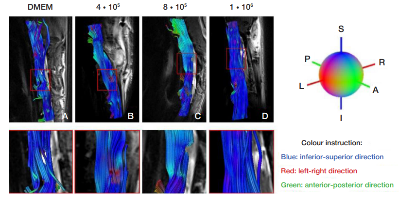
Fig. 4. Tractography images of the spinal cords in all the groups at 8w post cell transplantation. Tractography image of the spinal cord in DMEM group showed the fracture of neural fibers (A). All cell transplanted groups showed the repairing sign of spinal neural fibers (B–D). 1 • 106 group showed the best outcome (D)
Table 1. The cases counting of PTS formation
Table 2. The situation of ADC and FA in the injury area