METHOD

Cyclodialysis ab externo with implantation of a collagen implant in surgical management of glaucoma

About authors

1 Peoples’ Friendship University of Russia, Moscow, Russia

2 Eye microsurgery center «Pro zrenie», Moscow province, Russia

3 Ophthalmic unit of Skhodnya City Hospital, Khimki, Moscow province, Russia

Correspondence should be addressed: Ahmad S. Shradqa
proezd Shokalskogo, 13, bl. 1, Moscow, 127221; moc.liamg@wocsom8891hs

About paper

Author contribution: Shradqa AS — study concept and design, data collection and processing, statistical processing, article authoring, design of graphs and drawings; Kumar V — study concept and design, data collection and processing, statistical processing, article authoring and editing, overall responsibility; Frolov MA — editing and overall responsibility; Dushina GN — study concept and design, data collection and processing, article editing; Bezzabotnov AI — study concept and design; data collection and processing; Abu Zaalan KA — data collection and processing.
Received: 2019-08-16 Accepted: 2019-10-08 Published online: 2019-10-23
|
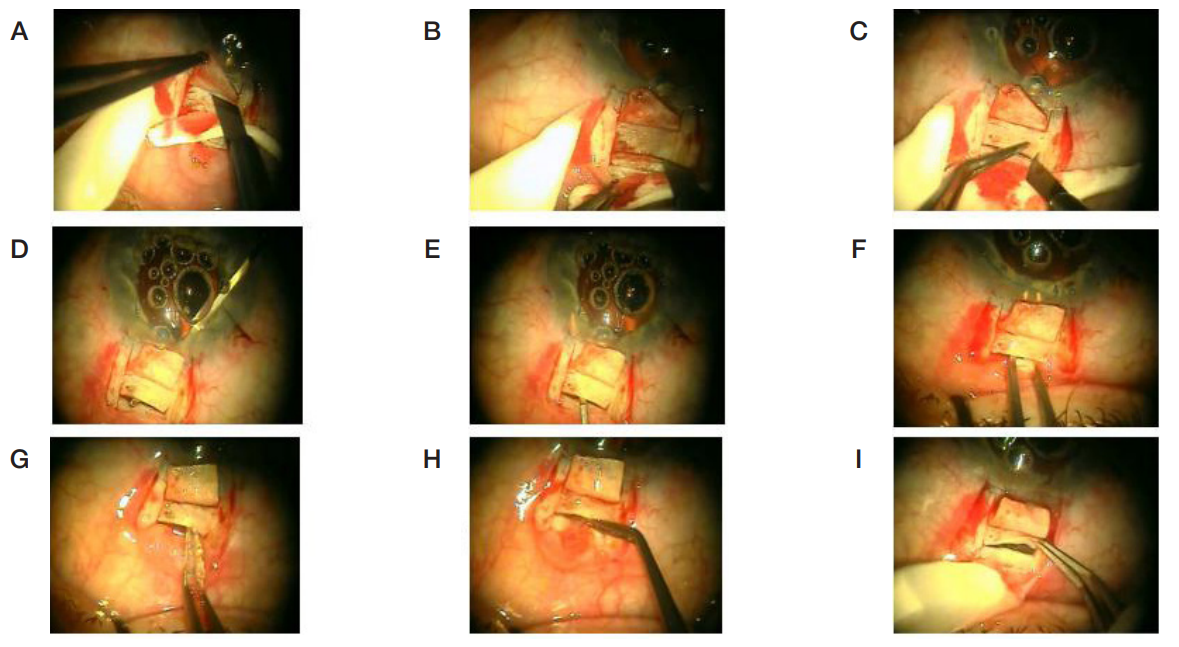
Fig. 1. A. Dissection of limbal based superficial scleral flap (3 × 2 mm). B. Horizontal incision of the sclera's deep layers up to the ciliary body. C. Formation of the valve by making 2 vertical incisions 1.0 mm long at both ends of the incision. D. Cyclodialysis ab interno step. Using a spatula inserted into AC through the paracentesis the ciliary body was separated from the scleral spur, spatula's distal end exiting through the incision in the sclera. E. Completion of tunnel formation ab externo. The spatula was inserted through the scleral incision, its end entered the AC; the ciliary body was detached from sclera along the entire length of the incision. F. CT expansion with the help of suture tying forceps. G. NACI implantation into the supraciliary space. Pre-moistened in the balanced salt solution NACI was captured between tying platforms of the forceps and was inserted into the tunnel through the incision in the sclera and was pushed forward till it appeared in the AC angle. H. NACI implantation in the suprachoroidal space. Posterior end of the implant was captured with toothed forceps and inserted under the back lip of the scleral incision into the suprachoroidal space. I. NACI position checkup
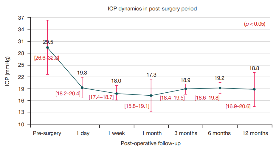
Fig. 2. Graph showing IOP dynamics after cyclodialysis ab externo with implantation of NACI at different follow-up visits
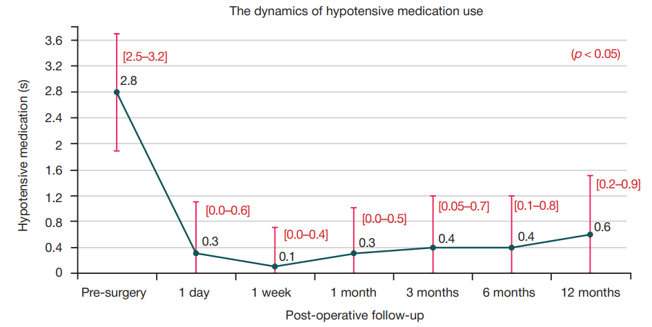
Fig. 3. Use of hypotensive medication by POAG patients at different follow-up visits
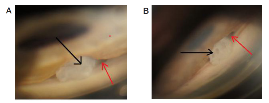
Fig. 4. Gonioscopy view of the AC angle. A. 1 month after surgery. B. 12 months after surgery. NACI (black arrow) is in the CT; CC is clearly visible (red arrow)
Fig. 5. Ultrasound examination of the surgical area. Post-operative follow-up — 12 months. A. Ultrasonic B-scan. The cyclodialysis tunnel is clearly visible. B. Ultrasonic biomicroscopy. The NACI is in the created tunnel. No dislocation was observed. Implants porous structure is clearly visible
Fig. 6. OCT of the anterior segment of the eye. Post-operative follow-up — 12 months. A. Transverse section. B. Iongitudinal section. The NACI in the supraciliary and suprachoroidal spaces is clearly visible. The spaces are expanded