ORIGINAL RESEARCH

Paraquat-induced model of Parkinson’s disease and detection of phosphorylated α-synuclein in the enteric nervous system of rats

About authors

Research Center of Neurology, Moscow, Russia

Correspondence should be addressed: Alla V. Stavrovskaya
Per. Obukha 5, Moscow, 103064; ur.liam@vats_alla

About paper

Funding: this work supported by the Russian Science Foundation (Grant 19-15-00320).

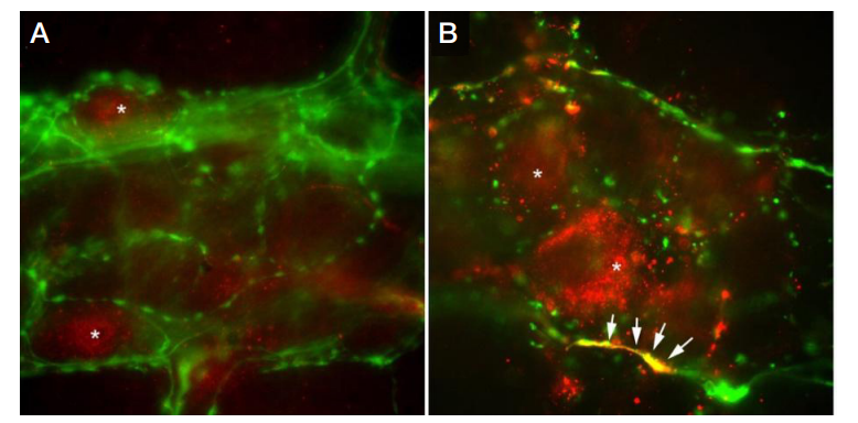
Acknowledgement: the authors thank their colleagues, Olshansky AS and Yamshchikova NG (the Laboratory of Experimental Pathology of the Nervous System), for their valuable contribution.

Author contribution: Stavrovskaya AV planned the study, analyzed the literature, collected, analyzed and interpreted the obtained data, conducted behavioral tests, administered drugs to the animals, and prepared the draft of the manuscript; Voronkov DN analyzed the literature, analyzed and interpreted the obtained data, prepared brain slides, conducted the histopathologic examination, and prepared the draft of the manuscript; Kutukova KA analyzed the literature, analyzed the obtained data, prepared jejunum slides, carried out the histopathologic examination, and prepared the draft of the manuscript; Ivanov MV prepared jejunum slides and carried out the histopathologic examination; Gushchina AS collected data, administered drugs to the rats, carried out behavioral tests, and monitored the animals’ health as a vet; Illarioshkin SN supervised the study and prepared the draft of the manuscript.

Received: 2019-08-12 Accepted: 2019-08-26 Published online: 2019-09-14
|
Fig. 1. The distance covered by the rats from the main and control groups in the open field test
Fig. 2. The number of slips made by the animals in the main and control groups during the tapered beam walking test expressed as % from the total number of steps (* — p = 0.0445)
Fig. 3. Changes in the striatum of the experimental animals following long-term exposure to paraquat. Reduced density of TH-positive fibers and astrocyte hypertrophy in the striatum of the control (A) and the main (B) groups. Staining for TH is shown in green; staining for GFAP, in red; DAPI staining, in blue (immunofluorescent staining; magnification ×40). C. Changes in the intensity of TH-positive staining expressed as % from the staining intensity observed in the intact controls (* — p = 0.033; Mann-Whitney U)
Fig. 4. Nervous fibers in the myenteric plexus of the rat jejunum containing TH (А) and class III β-tubulin (C) and changes in the fluorescence intensity of TH-positive (B) and class III β-tubulin-positive (D) fibers in the rats exposed to paraquat (% from the intensity observed in the control group; magnification ×10; * — p = 0.01)
Fig. 5. Location of α-Syn-p129 (shown in red) and TH (shown in green) in the myenteric plexus of the control group (A) and the rats exposed to paraquat (B). Synucleinpositive inclusions in TH-positive nervous fibers are marked by arrows; neuronal bodies are marked by asterisks (*) (magnification ×40)