ORIGINAL RESEARCH

Internal carotid and vertebral artery dissection: morphology, pathophysiology and provoking factors

Kalashnikova LA, Gulevskaya TS, Sakharova AV, Chaykovskaya RP, Gubanova MV, Danilova MS, Shabalina AA, Dobrynina LA
About authors

Research Center of Neurology, Moscow, Russia

Correspondence should be addressed: Ludmila A. Kalashnikova
Volokolamskoe shosse, 80, Moscow, 125367; ur.xednay@ncnavokinhsalak

About paper

Funding: this study was part of the state assignment for Research Center of Neurology.

Author contribution: Kalashnikova LA, Gubanova MV — literature analysis, data acquisition, processing of the obtained data, manuscript preparation; Gulveskaya TS, Sakharova AV, Chaykovskaya RP, Shabalina AA — data acquisition, analysis and interpretation; Danilova MS — recruitment of participants; Dobrynina LA — processing of the obtained data, manuscript preparation.
Received: 2019-09-03 Accepted: 2019-10-19 Published online: 2019-10-04
|
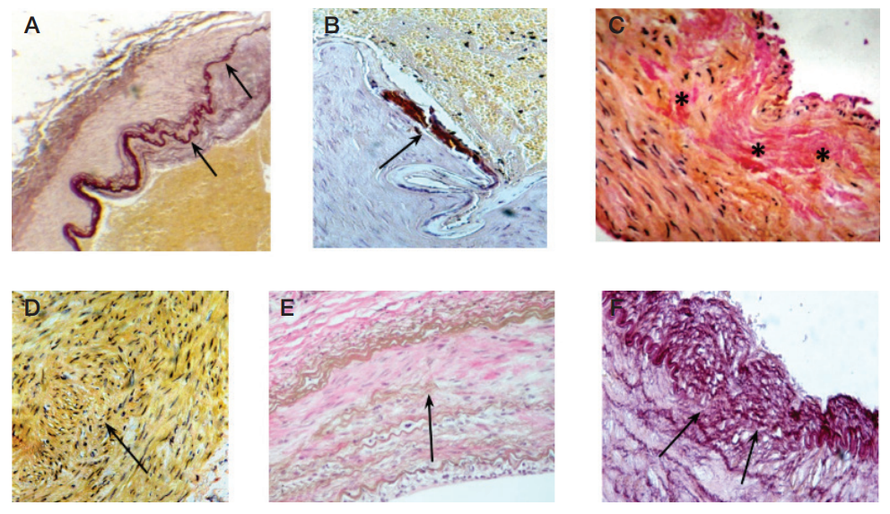
Figure. Arterial wall dysplasia in the internal carotid and vertebral arteries. A. Thinning and splitting of the internal elastic membrane in the dissected ICA siphon (shown by arrows). Staining: Weigert’s elastic stain (fuchselin); x100 magnification. B. Calcium deposits on the internal elastic membrane in the dissected ICA siphon (shown by an arrow). Staining: von Kossa method; x200 magnification. C. Areas of fibrosis (marked by an asterisk) in the tunica media of the extracranial segment of the dissected ICA. Staining: van Gieson’s stain; x200 magnification. D. An area of the tunica media in the dissected ICA siphon containing irregularly oriented myocyts (marked by an arrow). Staining: van Gieson’s stain; x200 magnification. E. Areas of fibrosis and the decreased number of elastic fibers in the tunica media of the intact internal carotid artery (shown by an arrow). Staining: van Gieson’s stain; x200 magnification. F. A fragment of the internal elastic membrane is missing (shown by an arrow) in the intact vertebral artery. Staining: Weigert’s elastic stain (fuchselin); x200 magnification
Table 1. CTD biomarkers in the patients with ICA/VA dissections in the controls (one-way ANOVA)
Note: p — statistical significance; SD — standard deviation.
Table 2. CTD biomarkers in patients with different localization of arterial dissection (one-way ANOVA and post-hoc tests + Scheffe's procedure)
Note: p — statistical significance; ICA — internal carotid artery; VA — vertebral artery; SD — standard deviation.