CLINICAL CASE

Case report: removal of a proliferating pilomatricoma with a CO2 laser

About authors

Pirogov Russian National Research Medical University, Moscow, Russia

Correspondence should be addressed: Tatiana A. Gaydina
Ostrovityanova, 1, Moscow, 117997; ur.xednay@924cod

About paper

Author contribution: Gaydina TA — surgery; literature analysis; data acquisition, analysis and interpretation; manuscript preparation; Dvornikov AS — literature analysis; data acquisition, analysis and interpretation; Skripkina PA — manuscript preparation.
Received: 2019-11-14 Accepted: 2019-12-21 Published online: 2019-12-01
|
Fig. 1. A pale-pink skin nodule with yellow inclusions on the left cheek
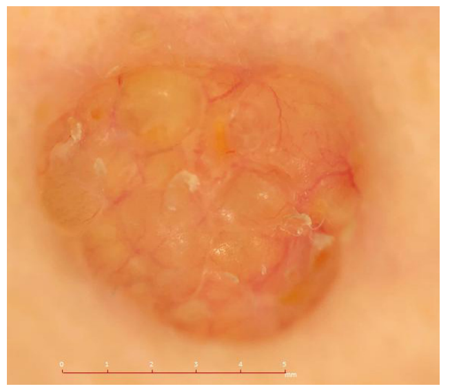
Fig. 2. Dermoscopy: a mass with a clear lobular structure, showing homogenous areas of yellow and pale pink, tortuous blood vessels and skin flakes (magnification: ×20)
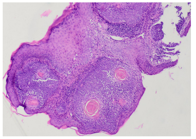
Fig. 3. A skin fragment with a nodule consisting of proliferating epithelial cells in a garland-resembling arrangement. Some of the cells are necrotic (the socalled shadow cells). Inflammatory cell infiltrate is seen around the layers of the epithelium (staining: hematoxylin and eosin; magnification ×40)
Fig. 4. Dermoscopy: a well-demarcated symmetrical pale pink area (magnification: ×20)
Fig. 5. A neat postoperative scar is staring to form