ORIGINAL RESEARCH

Rehabilitation of patients with cerebral palsy using hand exoskeleton controlled by brain-computer interface

Bobrov PD1,2, Biryukova EV1,2, Polyaev BA1, Lajsheva OA1,3, Usachjova EL3, Sokolova AV3, Mikhailova DI3, Dement'eva KN3, Fedotova IR2
About authors

1 Pirogov Russian National Research Medical University, Moscow, Russia

2 Institute of Higher Nervous Activity and Neurophysiology, Russian Academy of Sciences, Moscow, Russia

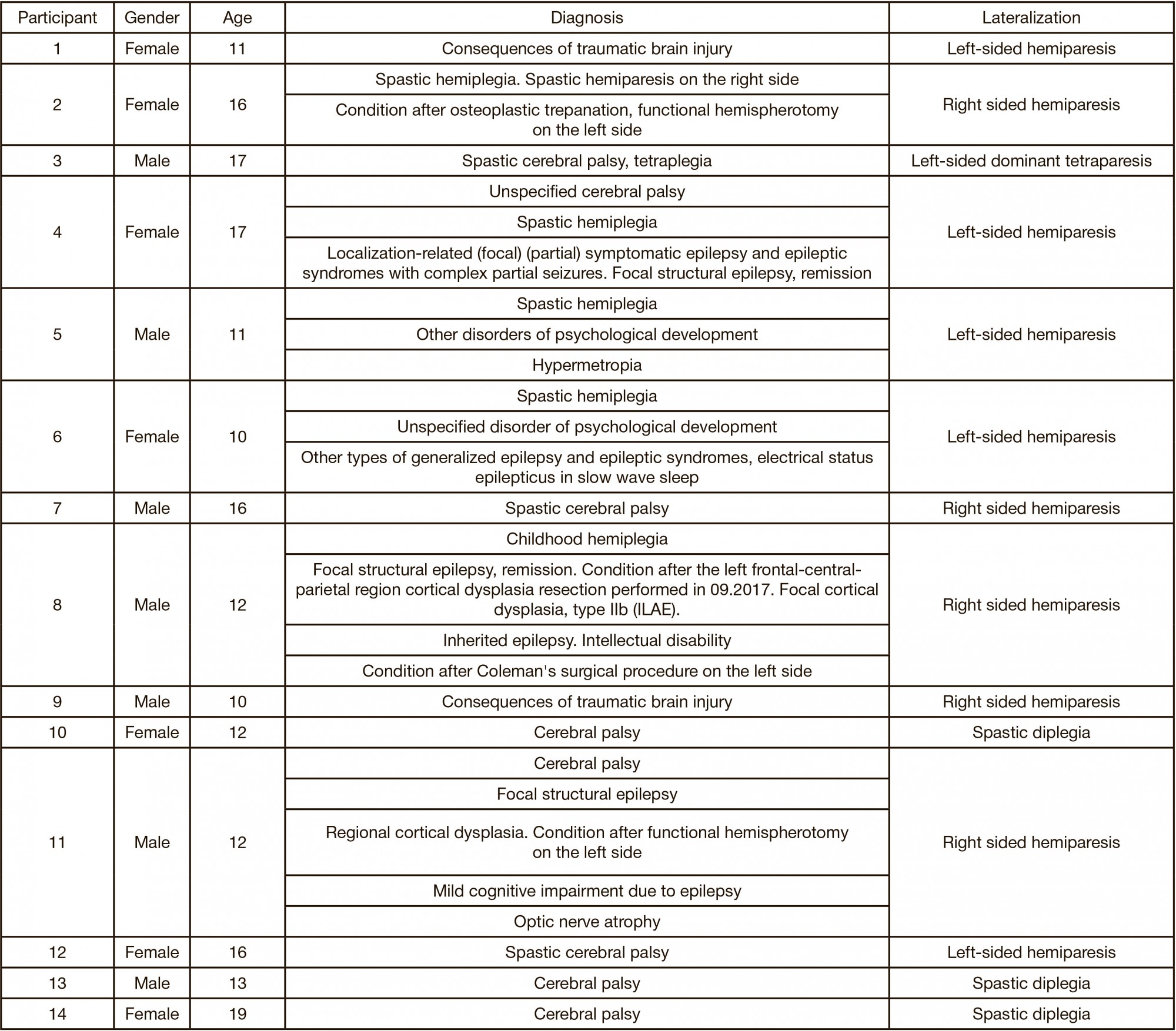
3 Russian Children's Clinical Hospital of Pirogov Russian National Research Medical University, Moscow, Russia

Correspondence should be addressed: Pavel D. Bobrov
Ostrovitianova, 1, Moscow, 117997; ur.xednay@vorbob-p

About paper

Funding: the study was supported by the Ministry of Science and Higher Education of the Russian Federation, project № RFMEFI60519X0184.

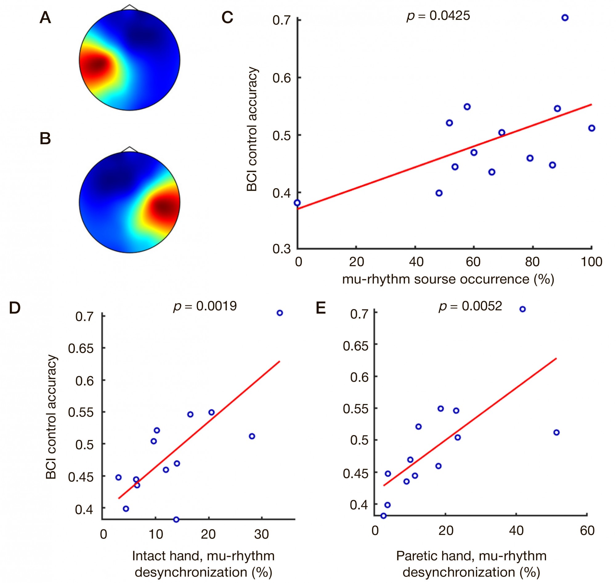
Author contribution: Bobrov PD — EEG processing and analysis, BCI accuracy estimation, manuscript writing; Biryukova EV — assessment scales scores statistical processing, manuscript writing; Polyaev BA, Lajsheva OA, Usachjova EL — clinical trial design; Usachjova EL — clinical trial management; Lajsheva OA, Sokolova AV, Mihailova DI, Dement’eva KN — development of methods for working with children, clinical data acquisition; Mihailova DI, Dement'eva KN — neuropsychological testing, training; Fedotova IR — literature analysis. All authors contributed to interpretation of the results and discussion.

Received: 2020-07-31 Accepted: 2020-08-13 Published online: 2020-08-20
|
Fig. 1. Topography of mu-rhythm sources and BCI control accuracy. А. Average topography of mu-rhythm sources in the left hemisphere. B. Average topography of mu-rhythm sources in the right hemisphere. C. Correlation between BCI control accuracy and mu-rhythm sources occurrence in all study participants. D. Correlation between BCI control accuracy and mu-rhythm desynchronization in the hemisphere opposite to the intact hand. E. Correlation between BCI control accuracy and murhythm desynchronization in the hemisphere opposite to the paretic hand
Table 1. Study participants’ characteristics
Table 2. Examples of clues simplifying the initiation of motor imagery
Table 3. Motor function median, quartile and significance level values according to Fugl-Meyer Assessment scale and ARAT test (obtained before and after the procedures)
Table 4. Motor function median, quartile (seconds) and significance level values according to Jebsen–Taylor function test (obtained before and after the procedures)