ORIGINAL RESEARCH

Efficacy of smartphone-compatible optical instrument for assessing melanocytic nevi for malignancy

Gaydina TA, Dvornikova EG
About authors

Pirogov Russian National Research Medical University, Moscow, Russia

Correspondence should be addressed: Tatiana A. Gaydina
Ostrovityanova, 1, Moscow, 117997; ur.xednay@924cod

About paper

Compliance with ethical standards: the study was approved by the Ethics Committee of Pirogov Russian National Research Medical University (Protocol № 201 dated October 21, 2020); all patients gave voluntary consent to surgery.

Author contribution: both authors equally contributed to this manuscript.
Received: 2020-09-11 Accepted: 2020-09-25 Published online: 2020-10-28
|
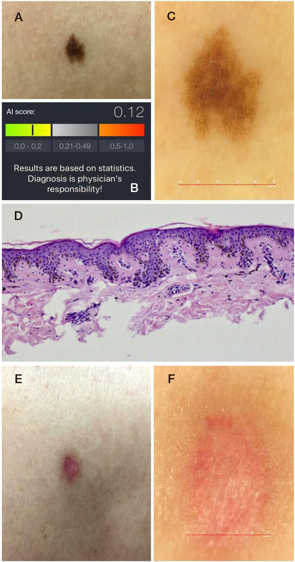
Fig. 1. AI in the assessment of MN for malignancy. A. A plain image of a melanocytic nevus. B. A malignancy score calculated by AI. C. A dermoscopy image at ×20 magnification: a reticular dysplastic nevus with hyperpigmentation at the center. D. A histological slide (hematoxilin-eosin stainin, ×100 magnification) showing the stratified squamous keratinized epithelium with areas of mild acanthosis. Multiple nevus cells adjacent to the epithelium are organized in a nesting pattern. Impression: D22. Borderline melanocytic nevus. ICD-O code: 8740/0. No nevoid cells are found at the margin of excision. D. Formation of a normotrophic scar. Е. A dermoscopy image of the normotrophic scar (at ×20 magnification)
Fig. 2. A. A preoperative photo of MMN on the abdomen. B. Postoperative scars