CLINICAL CASE

Staged approach to treatment of combined hamartoma of the retina and retinal pigment epithelium

About authors

Pirogov Russian National Research Medical University, Moscow, Russia

Correspondence should be addressed: Ekaterina P. Tebina
Volokolamskoe shosse, 30, str. 2, Moscow, 123182; ur.liam@anibetaniretake

About paper

Compliance with ethical standards: the patient submitted informed consent to staged surgery and personal data processing.

Author contribution: Takhchidi KhP — study concept and design, manuscript editing; Takhchidi NKh — literature analysis; Tebina EP — data acquisition and processing, manuscript writing; Kasminina TA — laser treatment.
Received: 2020-09-07 Accepted: 2020-10-03 Published online: 2020-10-12
|
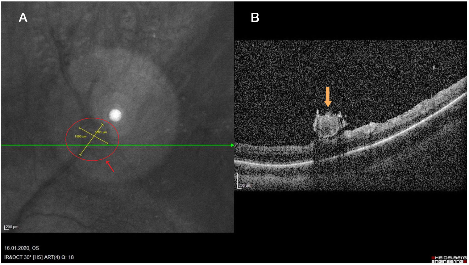
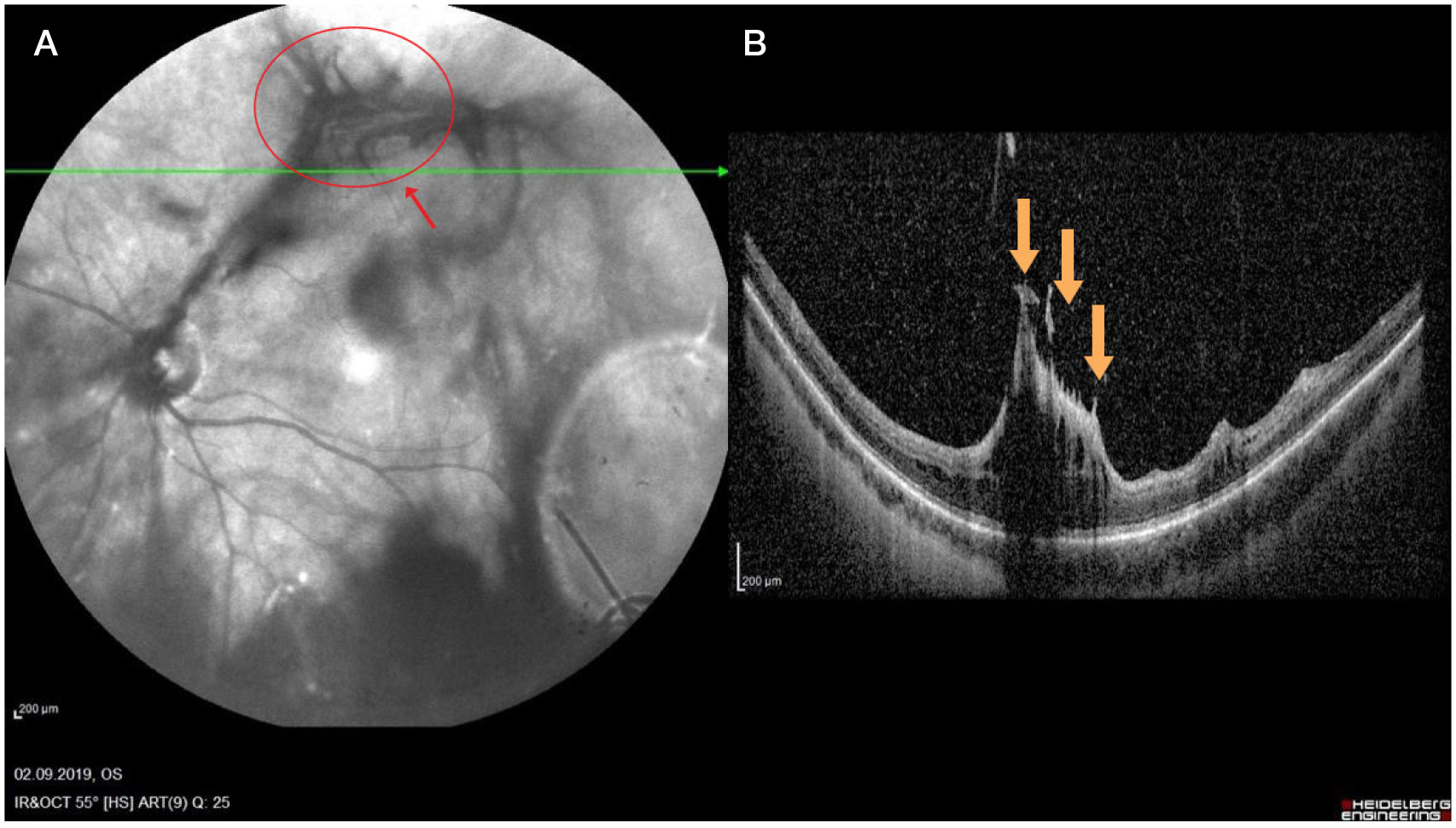
Fig. 1. А. IR reflectance image of fundus: protruding grey lesion with multiple sites of vitreoretinal traction in the area of the central retinal vein superior temporal branch. Partial hemophthalmos. B. OCT scan of retina: disorganization of the inner retinal layers in the protruding lesion area (maxi peaks, yellow arrow)
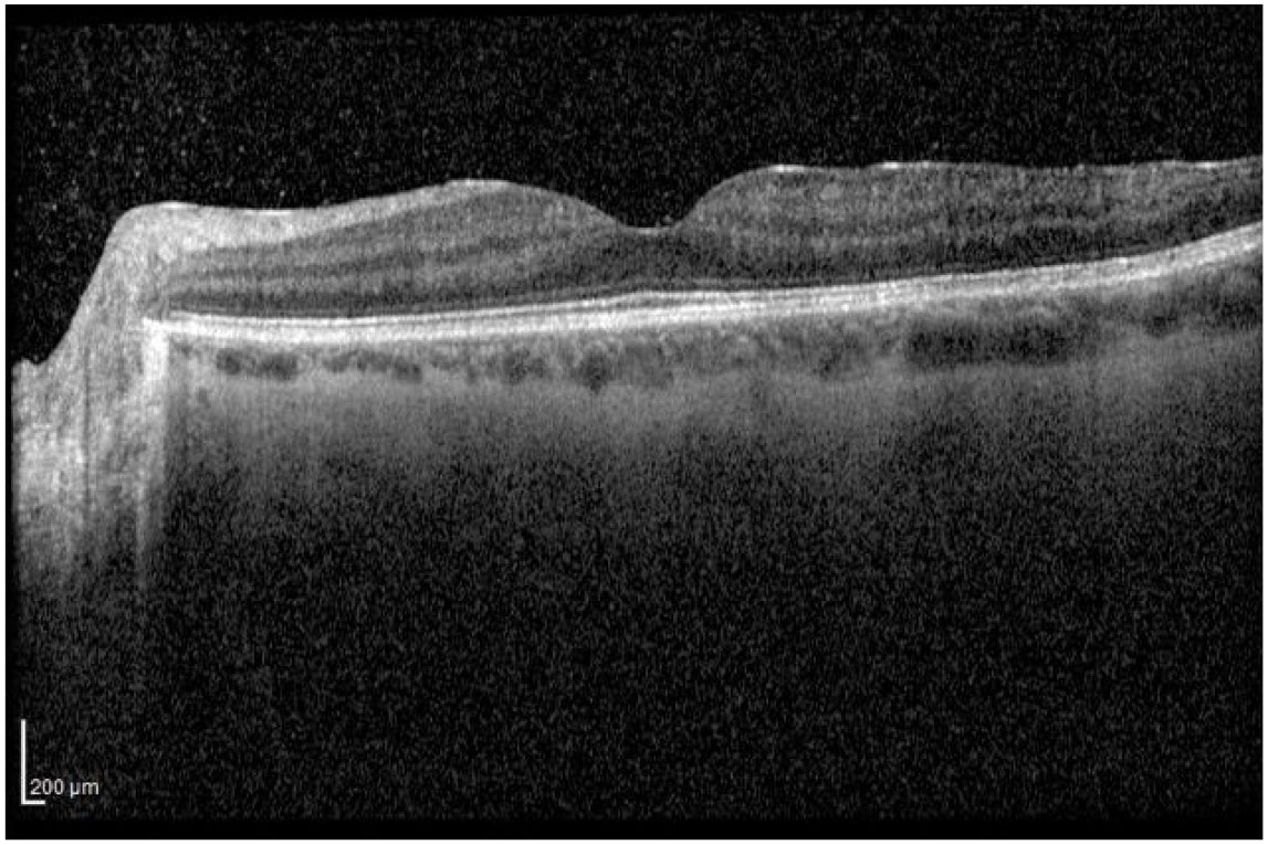
Fig. 2. OCT scan of retina: hyperreflective band in the central area of retinal surface, which is tightly attached to the internal limiting membrane, normal macular profile
Fig. 3. А. IR reflectance image of fundus a week after the subtotal vitrectomy: restored blood vessels calibre in the superior temporal branch, relieved vitreoretinal traction, shrunk protruding lesion (optic disk d = 0.5; red arrow) B. OCT scan of retina: single protruding lesion on the retinal surface (yellow arrow) with no vitreoretinal traction, restored anatomic structure of retinal layers