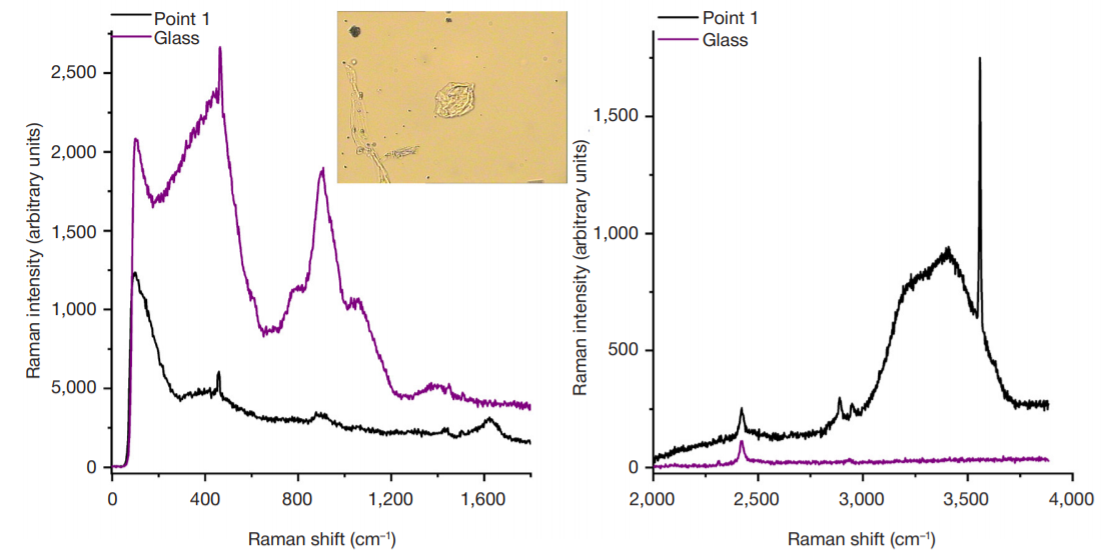
METHOD

Molecular and cellular features of mandibular autografts studied using raman spectroscopy

About authors

1 Lomonosov Moscow State University, Moscow, Russia

2 Pirogov Russian National Research Medical University, Moscow, Russia

3 A. I. Yevdokimov Moscow State University of Medicine and Dentistry, Moscow, Russia

4 National University of Science and Technology MISIS, Moscow, Russia

5 Central State Medical Academy of Department for Presidential Affairs of the Russian Federation, Moscow, Russia

Correspondence should be addressed: Georgy V. Maksimov
Leninskie Gory, 1, str. 24, Moscow, 117042; ur.liam@vomiskamg

About paper

Author contribution: Maksimov GV — study planning, analysis of the results; Sashkina TI — study planning, literature analysis; Fashutdinov DK — data acquisition and analysis, performing bone grafting; Slatinskaya OV — data processing; Saldusova IV — data analysis; Zaychenko OV — technical support.

Compliance with ethical standards: the study was approved by the Ethics Committee of the Central State Medical Academy of Department for Presidential Affairs of the Russian Federation (protocol № 3 dated September 23, 2021); the informed consent was submitted by all participants; biomaterials were treated in accordance with the World Medical Association Declaration of Helsinki.

Received: 2021-05-13 Accepted: 2021-05-31 Published online: 2021-06-17
|
Fig. 1. Raman spectrum of the autograft, sample № 2 (bone tissue and tissue components). The insert in the chart illustrates the image of the sample. The control is the Raman spectrum of the glass, the sample is placed on. X-axis: Raman shift (cm–1); Y-axis: Raman intensity (in arbitrary units)
Fig. 2. Raman spectrum of the autograft, sample № 2 (autograft tissues). The insert in the chart illustrates the image of the sample. The control is the Raman spectrum of the glass, the sample is placed on. X-axis: Raman shift (cm-1); Y-axis: Raman intensity (in arbitrary units)
Fig. 3. Raman spectrum of the autograft, sample № 1 (single cell). The insert in the chart illustrates the image of the sample. The control is the Raman spectrum of the glass, the sample is placed on. X-axis: Raman shift (cm-1); Y-axis: Raman intensity (in arbitrary units)
Fig. 4. Raman spectrum of the autograft, sample № 1 (red blood cell). The insert in the chart illustrates the image of the sample. The control is the Raman spectrum of the glass, the sample is placed on. X-axis: Raman shift (cm-1); Y-axis: Raman intensity (in arbitrary units)
Fig. 5. Raman spectrum of the autograft, sample № 2 (cell). The insert in the chart illustrates the image of the sample. The control is the Raman spectrum of the glass, the sample is placed on. X-axis: Raman shift (cm-1); Y-axis: Raman intensity (in arbitrary units)
Table. Studied samples