CLINICAL CASE

Long-term effects of multimodality laser therapy in patient with drusenoid pigment epithelial detachment

About authors

Pirogov Russian National Research Medical University, Moscow, Russia

Correspondence should be addressed: Nadezhda A. Mahno
Volokolamskoe shosse, 30, korp. 2, Moscow, 123182, Russia; moc.liamg@7onham.adzedan

About paper

Author contribution: Takhchidi KhP — study concept and design, manuscript editing; Takhchidi NKh — literature analysis; Kasmynina TA — laser therapy; Mahno NA — data acquisition and processing, manuscript writing.

Compliance with ethical standards: the patients submitted the informed consent to laser therapy and personal data processing.
Received: 2021-11-24 Accepted: 2021-12-16 Published online: 2021-12-29
|
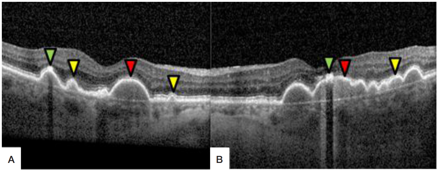
Fig. 1. Spectral-domain optical coherence tomography image of the fovea. A. Right eye: RPE detachment (red arrow) with the height of 166 μm and the length of 1126 μm, with homogeneously hyperreflective deposits. Coalescent soft drusen (yellow arrow), accumulation of subretinal hyperreflective deposits (green arrow). B. Left eye: RPE detachment (red arrow) with the height of 173 μm and the length of 2348 μm, with homogeneously hyperreflective deposits. Coalescent soft drusen (yellow arrow), accumulation of subretinal hyperreflective deposits (green arrow)
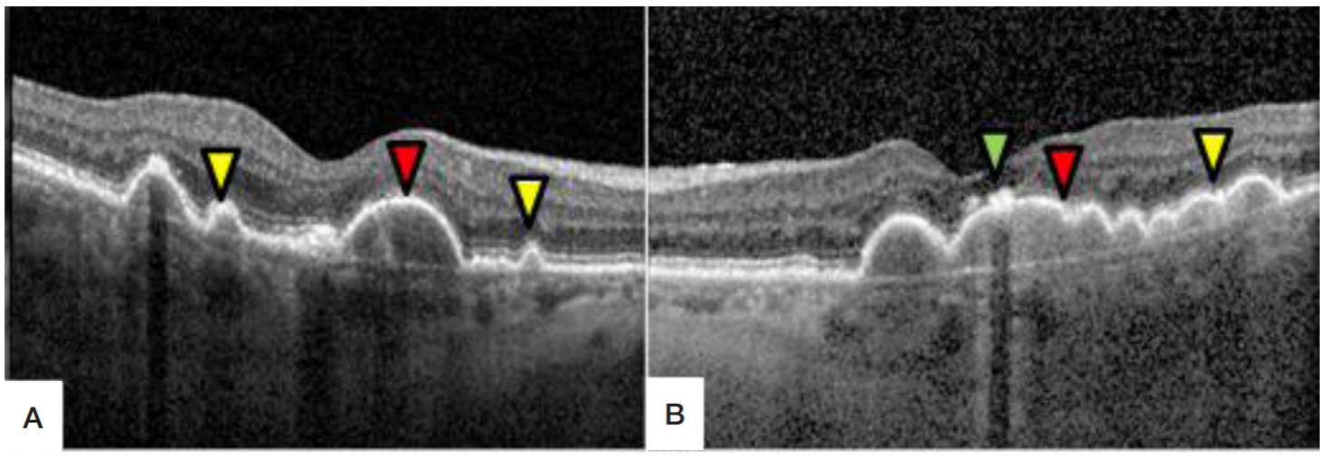
Fig. 2. Spectral-domain optical coherence tomography image of the fovea. A. Right eye: RPE detachment (red arrow) with the height of 164 μm and the length of 1081 μm, with homogeneously hyperreflective deposits. Coalescent soft drusen (yellow arrow), accumulation of subretinal hyperreflective deposits (green arrow). B. Left eye: RPE detachment with the height of 154 μm and the length of 2286 μm, with homogeneously hyperreflective deposits. Coalescent soft drusen (yellow arrow), accumulation of subretinal hyperreflective deposits (green arrow)
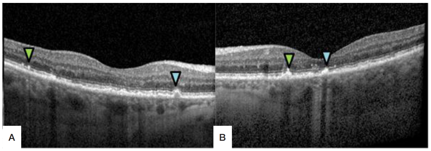
Fig. 3. Spectral-domain optical coherence tomography image of the fovea. A. Right eye: macula remains intact, undulating RPE line, sporadic hard drusen (blue arrow), accumulation of subretinal hyperreflective deposits (green arrow). B. Left eye: macula remains intact, undulating RPE line, sporadic hard drusen (blue arrow), accumulation of subretinal hyperreflective deposits (green arrow)
Fig. 4. Spectral-domain optical coherence tomography image of the fovea. A. Right eye: macula remains intact, undulating RPE line (orange arrow), sporadic hard drusen (blue arrow). B. Left eye: macula remains intact, undulating RPE line (orange arrow)