ORIGINAL RESEARCH

Ultrasound imaging of vagus nerves in patients with Parkinson's disease

About authors

Research Center of Neurology, Moscow, Russia

Correspondence should be addressed: Andrey О. Chechetkin
Volokolamskoe shosse, 80, Moscow, 125367, Russia; moc.liamg@niktehcehcyerdna

About paper

Author contribution: Chechetkin AO — study design, acquisition of ultrasound imaging data, data interpretation, manuscript preparation; Moskalenko AN — clinical data acquisition, analysis and interpretation; Fedotova EYu, Illarioshkin SN — study design, manuscript editing.

Compliance with ethical standards: the study was approved by the Ethics Committee of the Research Center of Neurology (Protocol No. 2-6/20 dated March 18, 2020)
Received: 2021-10-25 Accepted: 2021-11-11 Published online: 2021-11-24
|
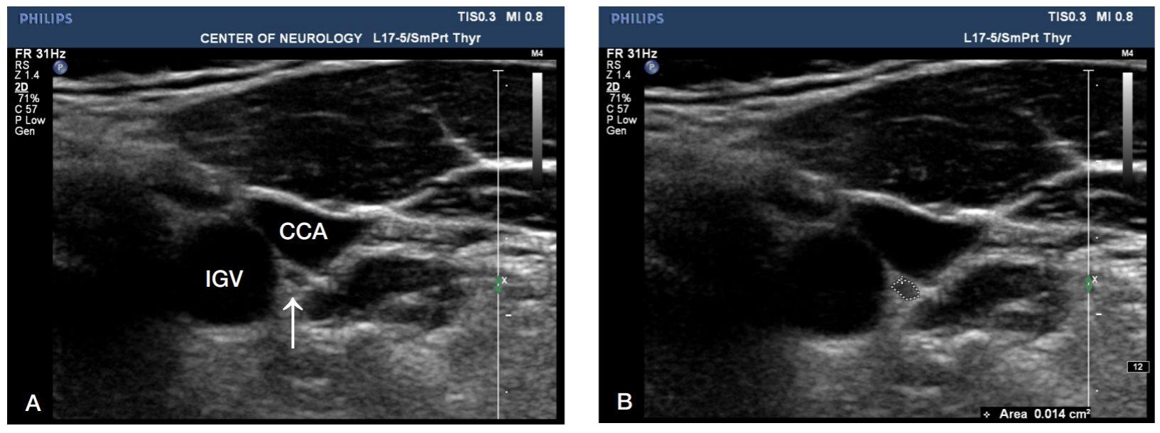
Fig. 1. A transverse ultrasound image of the vagus nerve. A. The left vagus nerve (indicated by the white arrow) is located between the common carotid artery (CCA) and the internal jugular vein (IGV). B. The same image showing delineation of the vagus nerve along the internal contour of the hyperechoic epineural rim. CSA (cross-sectional area) of the nerve = 0.014 cm2, or 1.4 mm2
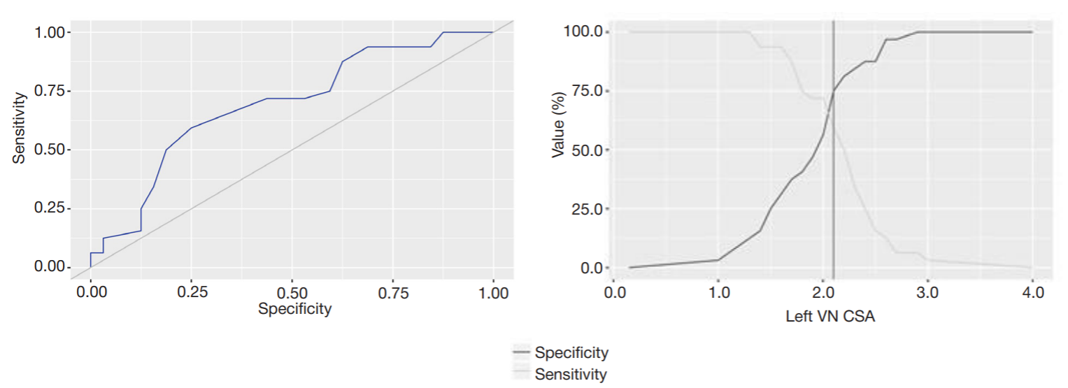
Fig. 2. Analysis of the model’s sensitivity and specificity depending on the threshold values of the left VN CSA. CSA — cross-sectional area; VN — vagus nerve
Fig. 3. The regression function graph for the association between the left VN CSA and SNH area on the right (A) and left (B). CSA — cross-sectional area; VN — vagus nerve; SNH — substantia nigra hyperechogenicity
Table 1. Clinical characteristics of patients with PD included in the study
Table 2. CSA of the right and left vagus nerves in patients with PD and healthy controls
Note: CSA — cross-sectional area; VN — vagus nerve; PD — Parkinson’s disease; * — p < 0.05, comparison with the control group.
Table 3. ЗVN CSA in patients with different forms of PD
Note: * — indicates statistically significant differences (p < 0.05).
Table 4. Ultrasonography findings in patients with PD
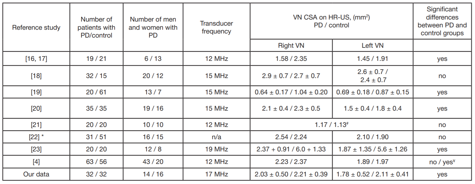
Note: PD — Parkinson’s disease; HR-US — highresolution ultrasound; CSA — cross-sectional area; VN — vagus nerve; * preprint, not peer reviewed yet; # — average diameter (mm); ¥ — reduction of right VN CSA in the PD group corrected for sex.