CLINICAL CASE

Laser treatment of macular retinal folds in late postoperative period after retinal detachment repair

About authors

Pirogov Russian National Research Medical University, Moscow, Russia

Correspondence should be addressed: Ekaterina P. Tebina
Volokolamskoe shosse, d. 30, korp. 2, Moscow, 123182, Russia; ur.liam@anibetaniretake

About paper

Author contribution: Takhchidi KhP — study concept and design, manuscript editing; Takhchidi EKh — literature analysis; Tebina EP — data acquisition, manuscript preparation; Kasminina TA — laser therapy.

Compliance with ethical standards: the patient gave informed consent to laser therapy and personal data processing.
Received: 2021-11-19 Accepted: 2021-12-05 Published online: 2021-12-21
|
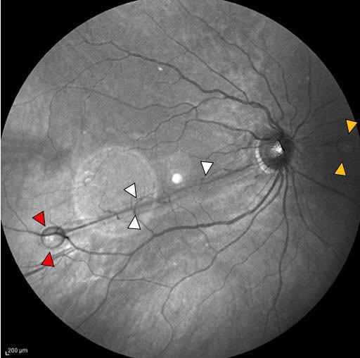
Fig. 1. The panoramic image of the right ocular fundus. A 756-µm PFCL bubble is visible peripapillarily on the nasal side (yellow arrow). A tight 633-µm-wide retinal fold extends from the temporal margin of the optic disc toward the 8 o’clock plane (white arrow) ending with a subretinal PFCL bubble sized 977 µm (red arrow). The fold involves half of the macular zone, predominantly its lower part
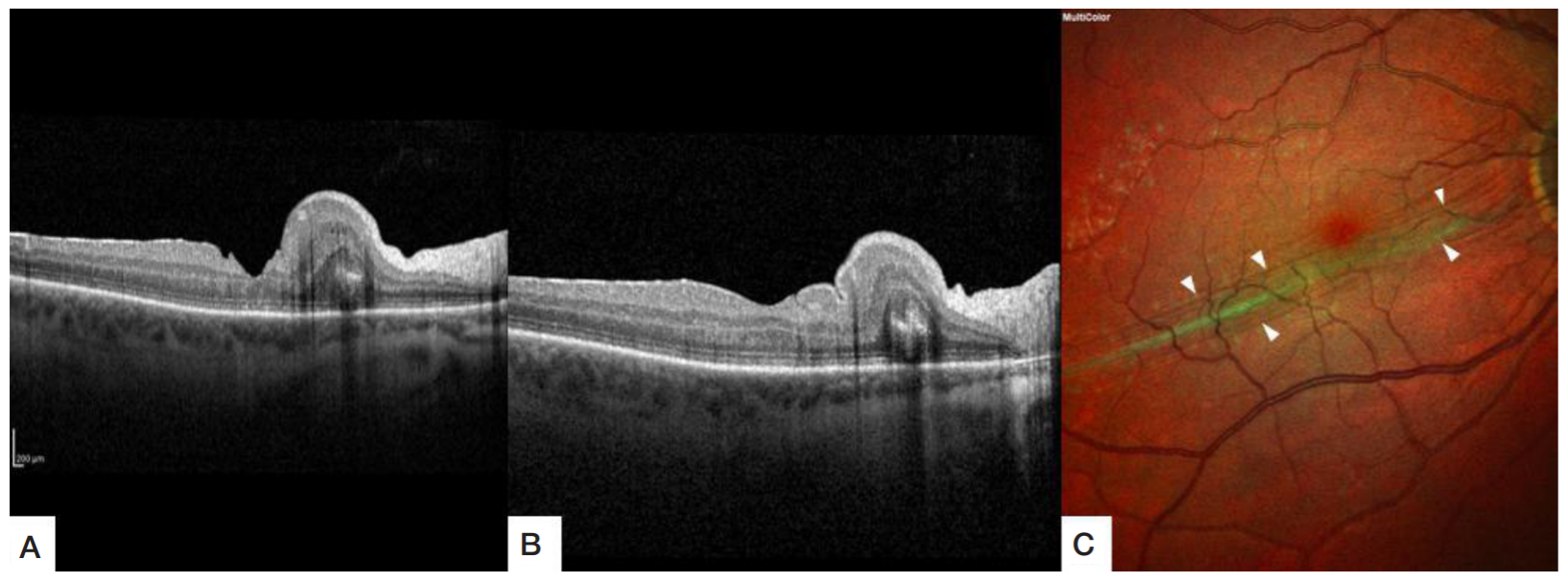
Fig. 2. Retinal spectral-domain optical coherence tomography scans of the right eye. A. The macula looks deformed, the foveal contour is normal (foveal depth: 185 µm, retinal thickness in the fovea: 192 µm); parafoveally, on the nasal side, the retina has a convex configuration showing a dominant retinal fold up to 637 µm in height, the retinal architecture looks grossly disorganized (changes involve the external limiting membrane, the connecting cilium between the inner and outer photoreceptor segments, the outer nuclear layer, and the outer plexiform layer). B. The macula is flattened; foveal depth: 106 µm, retinal thickness: 284 µm; a dominant full-thickness retinal fold up to 601 µm in height with gross retinal structure disorganization is visualized on the nasal side, further from the foveal margin. C. A multispectral image. Pronounced 633-µm-wide retinal folding extends from the temporal margin of the optic disc toward the 8 o’clock plane (white arrows)
Fig. 3. Retinal spectral-domain optical coherence tomography scans of the right eye. А. The macular profile is restored; the fovea is flattened: foveal depth 134 µm, retinal thickness in the fovea 240 µm; parafoveally on the nasal side, the retinal fold has diminished in height from 637 to 487 µm; the structural organization of the retinal layers appears partially restored; microfolds are visualized in the inner retinal layers paramacularly on the temporal side. B. The foveal contour is clear; foveal depth has increased to 142 µm, retinal thickness in the fovea has decreased to 243 µm; parafoveally on the nasal side, the retinal fold has diminished in height from 601 to 473 µm, the structural organization of the retinal layers looks partially restored. C. Infrared image. In the central zone, the folds have become less pronounced and flattened to 921 µm in width (white arrow)
Fig. 4. Retinal spectral-domain optical coherence tomography scans of the right eye. А. The macular profile is restored, the fovea is flattened; foveal depth has decreased to124 µm, retinal thickness in the fovea has increased to 273 µm; paramacularly, on the nasal side, intraretinal coagulation spots are visible at the RPE level (red arrow). В. The macular profile is restored, the fovea has a clear contour; foveal depth: 160 µm, retinal thickness in the fovea: 224 µm, the structural organization of the retinal layers is restored; paramacularly on the nasal side microfolds are visible in the inner retinal layers. C. A multispectral image: the retinal folds have unfolded (white arrow), weakly pigmented coagulation spots are visible in the macular zone, except for the avascular area, and in the epiretinal membrane area.