OPINION

Intermembrane oligomerization of SARS-CoV-2 M-protein: possible role in viral budding

About authors

Center for Molecular and Cellular Biology, Skolkovo Institute of Science and Technology, Moscow, Russia

Correspondence should be addressed: Konstantin A. Lukyanov
Bolshoy Bulvar, 30, str. 1, Moscow, 121205, Russia; moc.liamg@nitnatsnok.vonaykul

About paper

Funding: the study was funded by the Russian Foundation for Basic Research, project number 20-04-60370.

Author contribution: Sokolinskaya EL, Putlyaeva LV, Gorshkova AA — experiments; Lukyanov KA — concept and writing.
Received: 2022-05-13 Accepted: 2022-05-28 Published online: 2022-06-04
|
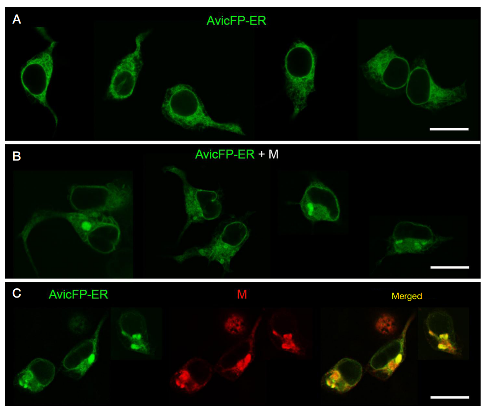
Fig. 1. Confocal fluorescence microscopy of HEK293T cells transfected with expression constructs. Scale bars, 20 µm. A. Transfection with plasmid encoding fluorescent protein AvicFP-ER localized to ER (green channel). B. Cotransfection with plasmids encoding AvicFP-ER (green channel) and M-protein. C. Cotransfection with plasmids encoding AvicFP-ER (green channel) and M-protein with subsequent immunostaining for M-protein (red channel; a merged channel image is shown on the right). The images are collages of four (A), four (B) and two (C) fields of view, showing representative results of each experiment.
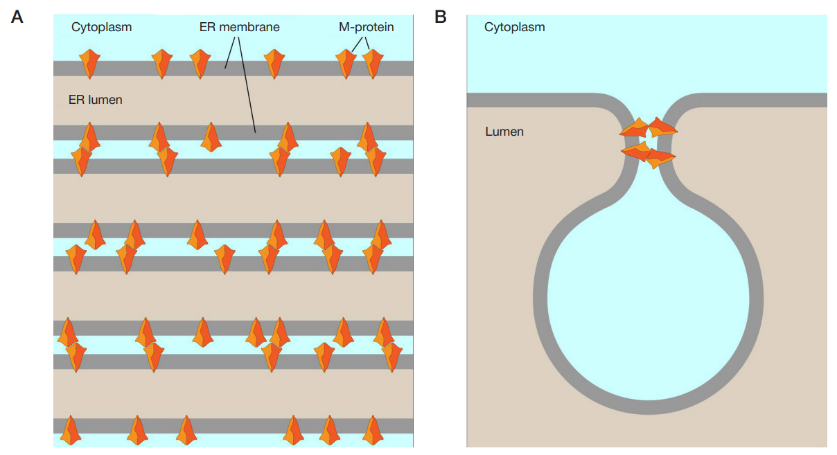
Fig. 2. A scheme of SARS-CoV-2 М-protein intermembrane oligomerization. A. Stacking of ER membranes into OSER structure through interactions of М-protein dimers in adjacent membranes. B. Hypothetical participation of the М-protein intermembrane oligomers in the neck formation during budding (М-protein molecules at other locations are omitted for clearance).