ISSN Print 2500–1094
ISSN Online 2542–1204
Bulletin of RSMU
BIOMEDICAL JOURNAL OF PIROGOV UNIVERSITY (MOSCOW, RUSSIA)
1 Belgorod State National Research University, Belgorod, Russia
2 Sechenov First Moscow State Medical University, Moscow, Russia
3 Kursk State Medical University, Kursk, Russia
4 Federal Research and Clinical Center of Intensive Care Medicine and Rehabilitology, Moscow, Russia
Correspondence should be addressed: Vladislav O. Soldatov
Pobedy, 85, Belgorod, 308015, Russia; moc.liamg@votadlosmrahp
Funding: State Assignment "Laboratory for Genome Editing in Biomedicine and Veterinary FZWG-2021-0016".
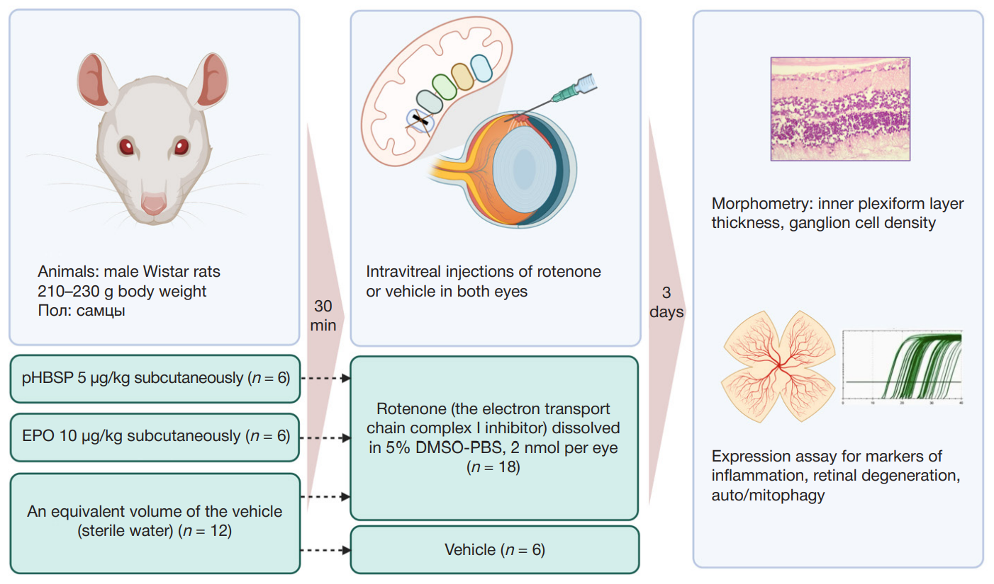
Author contribution: Soldatov VO — concept and design of the study, writing of the manuscript; Pokrovsky MV, Lapin KN — consulting on the concept and design of the study; Puchenkova OA — collection of samples for histological examination and target gene expression analysis; Puchenkova OA, Zhunusov NS — modeling of the rotenone-induced retinal degeneration; Zhunusov NS, Krayushkina AM — in vitro assay of rotenone cytoprotective activity; Grechina AV — morphological study; Bushueva OYu, Soldatova MO — RNA extraction, quantitative PCR assay.
Compliance with ethical standards: the study was approved by the ethical committee of the Belgorod State National Research University (Protocol № 06-07/21 of 15 July 2021); all procedures were performed in accordance with the Regulations on Laboratory Practice in the Russian Federation of 2003, in compliance with the 86/609 EEC Directive and ARRIVE guidelines.