ORIGINAL RESEARCH

Survival of human cells in tissue-engineered constructs stored at room temperature

Rogovaya OS1, Alpeeva EV1, Ruchko ES1, Eremeev AV1,2, Vorotelyak EA1
About authors

1 Koltzov Institute of Developmental Biology, Russian Academy of Sciences, Moscow, Russia

2 Lopukhin Federal Research and Clinical Center of Physical-Chemical Medicine of the Federal Medical Biological Agency, Moscow, Russia

Correspondence should be addressed: Olga S. Rogovaya
Vavilova, 26, Moscow, 119334, Russia; ur.xednay@f62ayavogor

About paper

Funding: the study was supported by the Ministry of Science and Higher Education of the Russian Federation, Agreement № 075-15-2021-1063 of 28.09.2021.

Author contribution: Rogovaya OS, Eremeev AV — experimental procedure, data analysis; Alpeeva EV — data interpretation, literature review; Ruchko ES — experimental procedure; Vorotelyak EA — study planning.

Compliance with ethical standards: the study was approved by the Ethics Committee of the Koltzov Institute of Developmental Biology, RAS (protocol № 51 of 09 September 2021) and conducted in accordance with the principles of the WMA Declaration of Helsinki and its subsequent revisions.
Received: 2022-12-16 Accepted: 2023-01-15 Published online: 2023-01-30
|
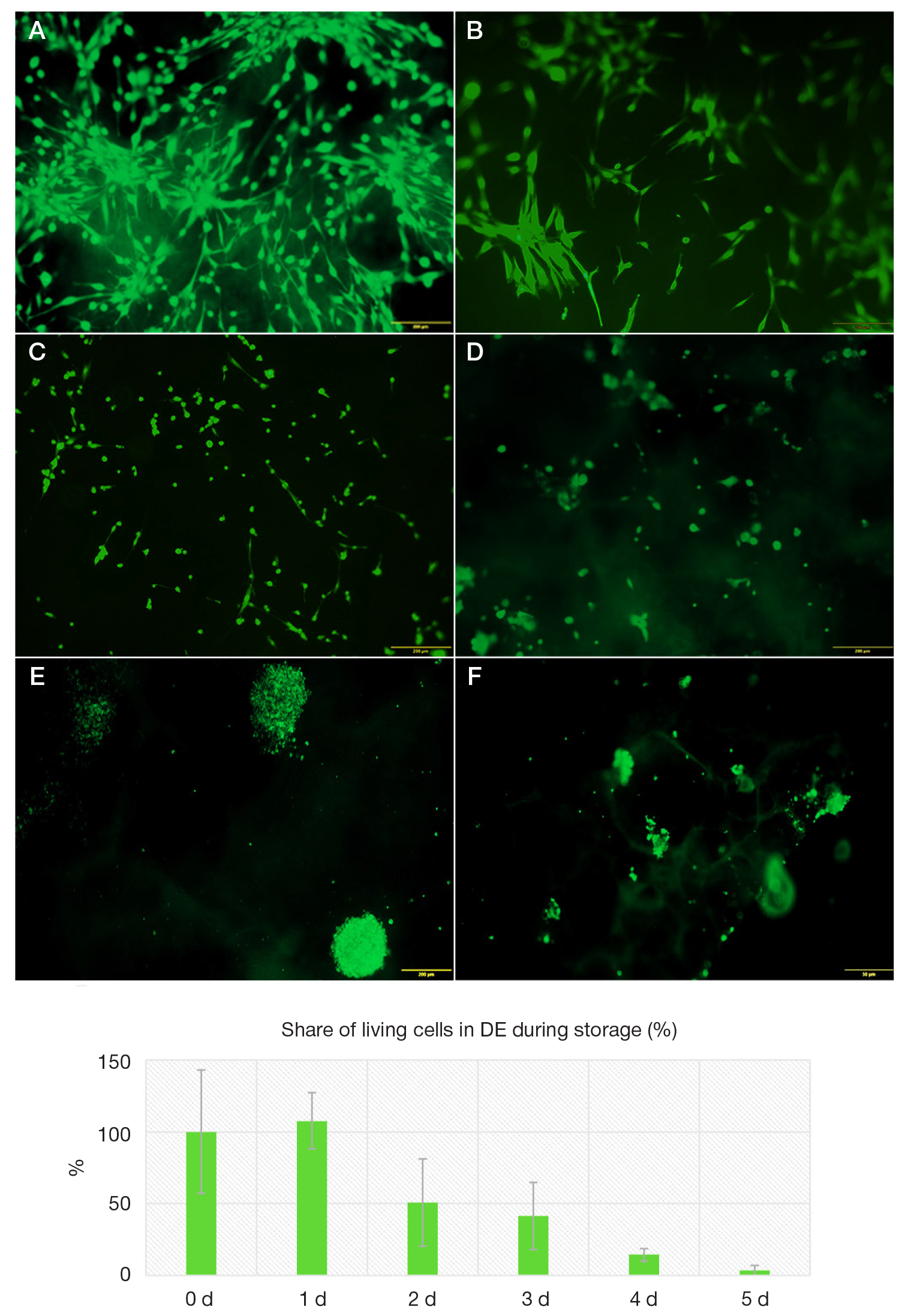
Fig. 1. Identification of living cells stained with the calcein vital dye (green stain) on the matrix surface in the DE TEC at various stages of storage at a temperature of +22 °C (А–F): at the beginning of the experiment (no storage, zero day) (А), day one (B), day two (C), day three (D), day four (E), and day five (F) of storage. G. The graph showing the percentage of living cells in the DE TEC as a function of the sample condition on the zero day of storage. The data are presented as the mean share of green pixels in three fields of view for three technical repetitions, error bars ±SD (%)
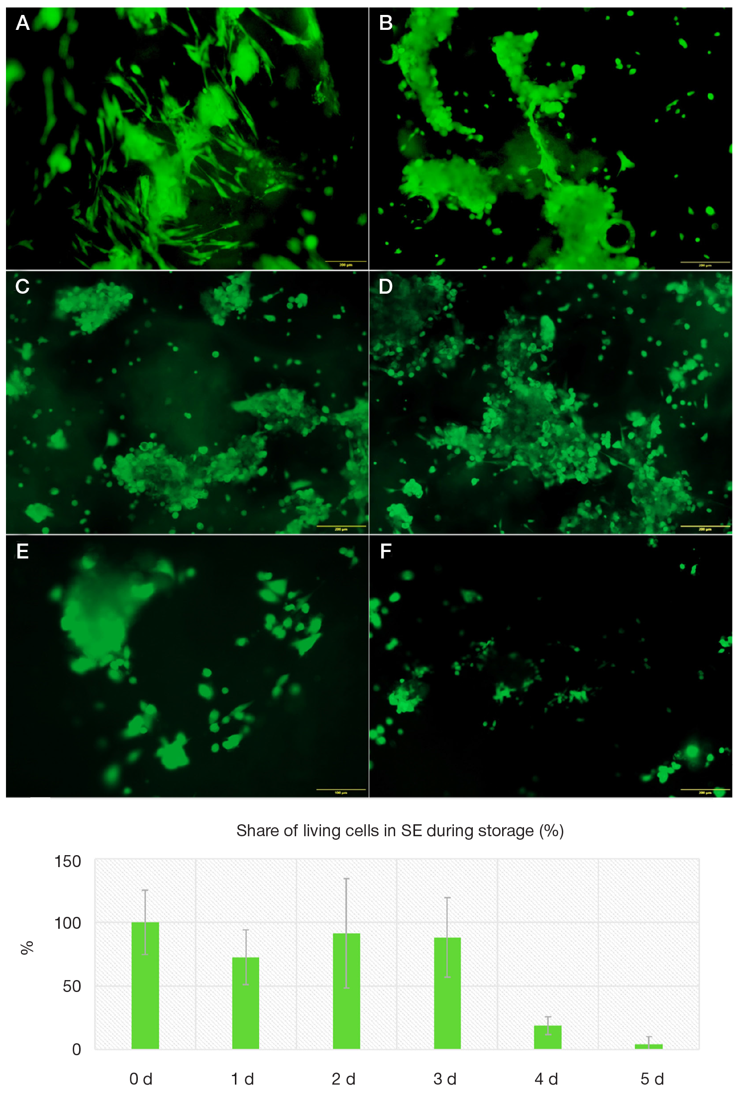
Fig. 2. Identification of living cells stained with the calcein vital dye (green stain) on the matrix surface in the SE TEC at various stages of storage at a temperature of +22 °C (А–F): at the beginning of the experiment (no storage, zero day) (А), day one (B), day two (C), day three (D), day four (E), and day five (F) of storage. G. The graph showing the percentage of living cells in the SE TEC as a function of the sample condition on the zero day of storage. The data are presented as the mean share of green pixels in three fields of view for three technical repetitions, error bars ±SD (%)
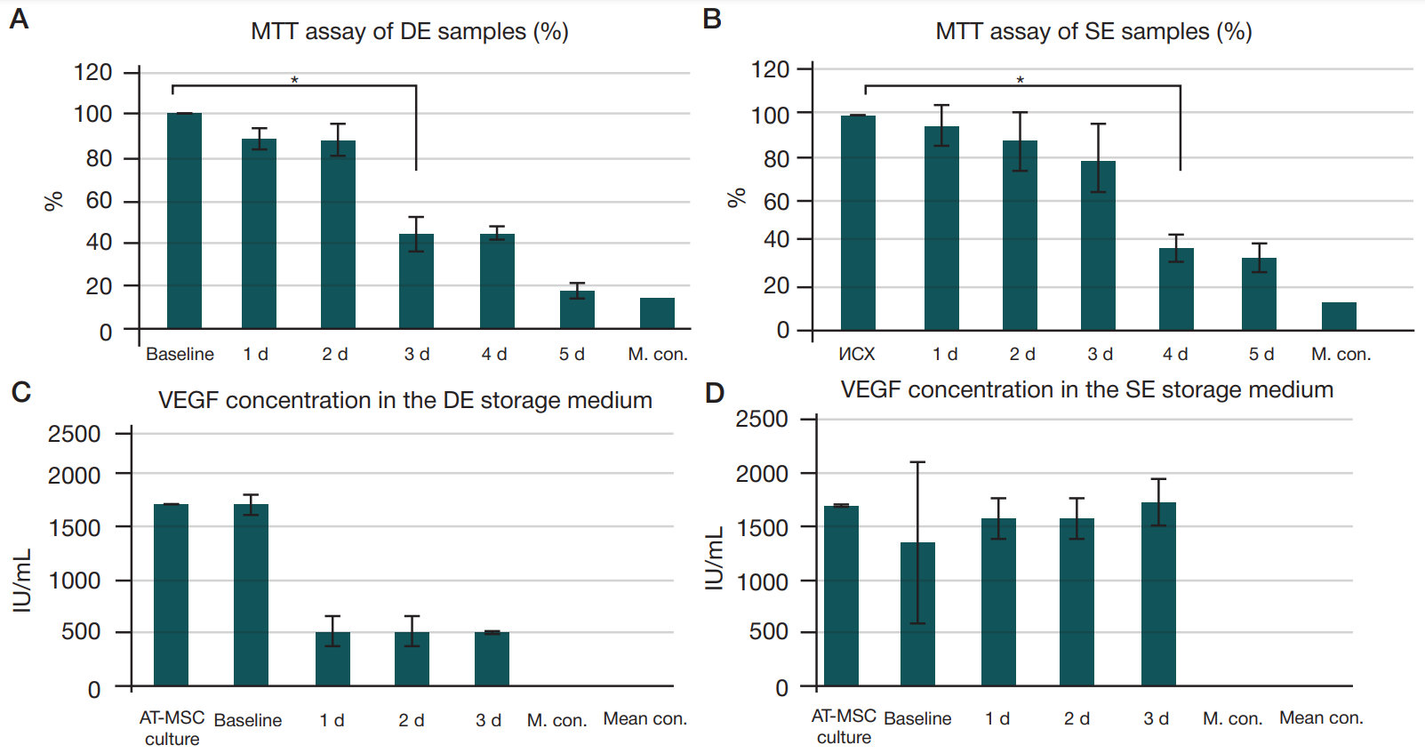
Fig. 3. The changes in the cells' mitochondrial activity assessed by MTT assay in the DE (А) and SE (B) TECs and presented as mean optical density values obtained at various stages of storage vs. data obtained before storage (zero day), percentage obtained in three technical repetitions, error bars ±SD (%). Significance of the metabolic activity decline observed on day three in DEs and day four in SEs is calculated by ANOVA (р ≤ 0.005). The data on the VEGF levels in the normal saline used for the DE (C) and SE (D) TEC sample storage measured by enzyme immunoassay are presented as mean optical density for nine repetitions, error bars ±SD (%). Matrix with no cells, culture medium, and original AT-MSC culture used to construct TECs were used as controls. There were no significant differences between the normal saline samples in the storage groups within three days
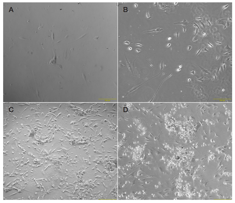
Fig. 4. The culture of cells extracted from DE (А, B) and SE (C, D) TECs after 72 h of storage. Cells after one day (А, C) and three days (B, D) after extraction (light microscopy, phase contrast)