CLINICAL CASE

Single-stage endovitreal surgery of retinal detachment complicated by macular hole involving the short-term perfluorocarbon tamponade

About authors

Pirogov Russian National Research Medical University, Moscow, Russia

Correspondence should be addressed: Nadezhda A. Mahno
Volokolamskoe shosse, 30/2, Moscow, 123182, Russia; moc.liamg@7onham.adzedan

About paper

Author contribution: Takhchidi KhP — study concept and design, surgical treatment of the patient, manuscript editing; Takhchidi NKh — literature review; Mahno NA — data acquisition and processing, manuscript writing.

Compliance with ethical standards: the patient submitted the informed consent to surgery and personal data processing.
Received: 2023-03-27 Accepted: 2023-04-21 Published online: 2023-04-28
|
Fig. 1. Spectral domain optical coherence tomography scan of the macular zone (before surgery). Retinal detachment. Macular hole (red arrow) with a diameter of 380 µm
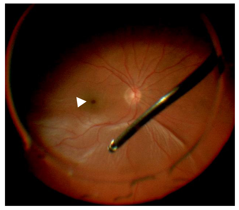
Fig. 2. Intraoperative fundus image: macular hole in the center of the retina (white arrow). Retinal detachment in the superior, temporal and inferior quadrants
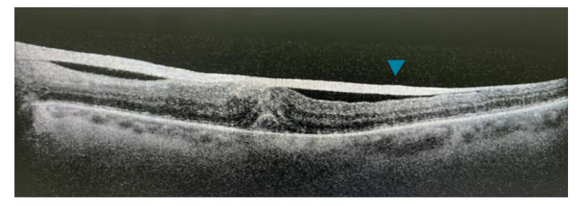
Fig. 3. Spectral domain optical coherence tomography scan of the macular region (24 h after surgery). The macular hole is closed, there is a hyperreflective structure represented by the fibrin film on the retinal surface (blue arrow)
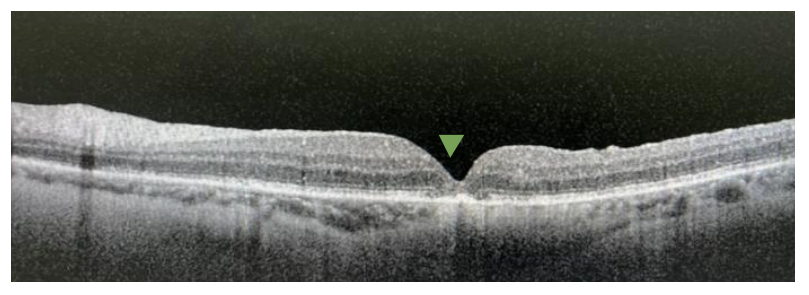
Fig. 4. Spectral domain optical coherence tomography scan of the macular region (one month after surgery). Apparent macular profile (green arrow). The “retinal pigment epithelium — Bruch's membrane” complex and discontinuous external limiting membrane are visible
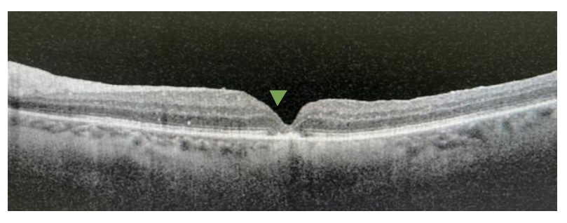
Fig. 5. Spectral domain optical coherence tomography scan of the macular region (six months after surgery). Apparent macular profile (green arrow). The “retinal pigment epithelium — Bruch's membrane”complex is visible. Partially restored segmentation of the external retinal layers (discontinuous external limiting membrane), partially restored photoreceptor zone of the retina