ORIGINAL RESEARCH

Preoperative planning of hip arthroplasty

Minasov BSh, Yakupov RR, Bilyalov AR, Minasov TB, Valeev MM, Mavlyutov TR, Nigamedzanov IE, Akbashev VN, Karimov KK
About authors

Bashkir State Medical University, Ufa, Russia

Correspondence should be addressed: Vladislav N. Akbashev
Lenina, 3, 45008, Ufa, Russia; ur.liam@bka-dalV

About paper

Funding: the study was supported by grant of the Government of the Republic of Bashkortostan for state support of scientific research guided by the leading scientists within the framework of the Eurasian Research and Educational Center programs; it was also supported by the Strategic Academic Leadership Program of the Bashkir State Medical University (PRIORITY-2030).

Author contribution: Minasov BSh, Yakupov RR, Bilyalov AR — developing the study design, data analysis; Minasov TB, Valeev MM, Mavlyutov TR — intraoperative control of determining the size of the endoprosthesis components, data acquisition, data analysis; Nigamedzanov IE, Akbashev VN, Karimov KK — statistical analysis, data estimation, literature review, computer, volumetric modeling, 3D printing of pelvic bones, acetabular and femoral components of the endoprosthesis.

Compliance with ethical standards: the study was approved by the Ethics Committee of the Bashkir State Medical University (protocol № 11 dated 15 November 2023)
Received: 2023-11-12 Accepted: 2023-12-19 Published online: 2023-12-31
|
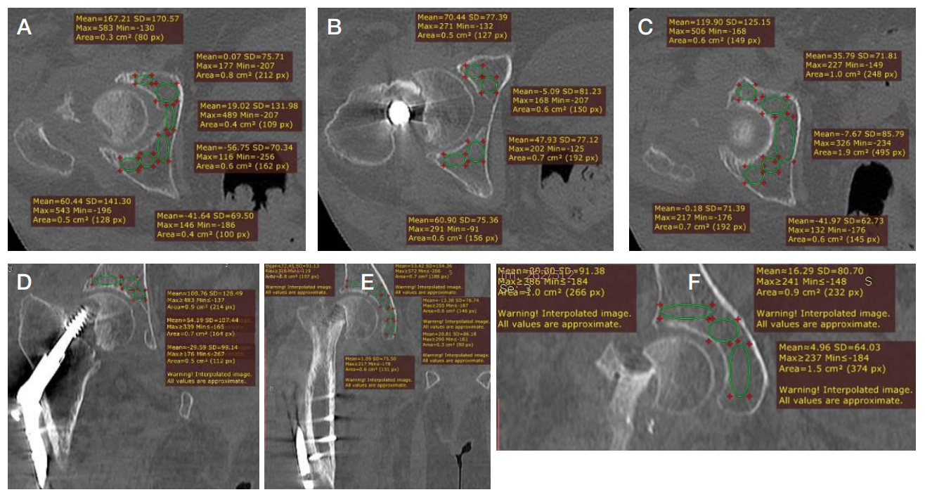
Fig. 1. Determination of bone density in the zones of fixation located in the acetabular area based on the horizontal (A, B, C) and frontal (D, E, F) CT scans of the 73-year-old patient: at the level of inferior acetabulum (А); at the level of the acetabulum middle part (B); at the level of superior acetabulum (C) (Average total bone density according to the Hounsfield scale — 30.65 HU.); at the level of posterior acetabulum (D); at the level of the acetabulum middle part (E); at the level of anterior acetabulum (F). (Average total bone density according to the Hounsfield scale — 30.09 HU.)
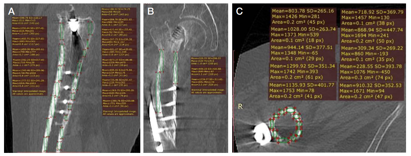
Fig. 2. Determination of bone density in the zones of fixation located in the acetabular area based on the sagittal, horizontal and frontal CT scans of the 73-year-old patient. А. Bone density estimation based on the sagittal scan. B. Bone density estimation based on the frontal scan. C. Bone density estimation based on the horizontal scan. (Average total bone density according to the Hounsfield scale — 869.13 HU.)
Fig. 3. Survey frontal x-ray image of the pelvis and the right hip joint of the 73-year-old patient. Diagnosis: condition after osteosynthesis involving the use of the DHS system. False joint of the right femoral neck. A. X-ray image acquired at admission to surgery — osteosynthesis of the right femur involving the use of the DHS system. B. X-ray image acquired after osteosynthesis involving the use of the DHS system. C. Sizing and positioning of the endoprosthesis components using the TraumaCad v. 2.4 software. (Planned size of the acetabular component — 50, femoral component — 4)
Fig. 4. Female patient, 73 years. Comparison of the endoprosthesis component dimensions during preoperative planning based on volumetric modeling performed before and after hip arthroplasty. A. Volumetric model of the right hip joint with virtual installation of the endoprosthesis components (planned size of the acetabular component — 52, femoral component — 6). B. Survey x-ray image of the pelvis and right hip joint after installation of endoprosthesis, the dimensions of acetabular and femoral components corresponded to the planned dimensions
Fig. 5. Patient, 45 years. Preoperative planning based on volumetric prototyping. A. Printed 3D model of the hip joint before arthroplasty. B. Printed 3D model of the hip joint after arthroplasty. C. Preoperative planning of the right hip arthroplasty based on volumetric modeling
Fig. 6. Implant sizing accuracy when using different preoperative planning methods
Table 1. Endoprothesis component sizing accuracy depending on the nosological form of degenerative and dystrophic disease of the hip joint (%)
Note: pc — significance of differences from controls.
Table 2. Selection of preoperative planning technique based on the form of degenerative and dystrophic disease of the hip joint