ORIGINAL RESEARCH

NEAT1_1 long non-coding RNA reduces the survival of primary neuronal cells under ER-stress

Pukaeva NE1,2, Zalevskaya VN2, Deykin AV3, Taubinskaya MI2, Kukharskaya OA2, Ovchinnikov RK1,2, Antohin AI1, Kukharsky MS1,2
About authors

1 Pirogov Russian National Research Medical University, Moscow, Russia

2 Institute of Physiologically Active Compounds at Federal Research Center of Problems of Chemical Physics and Medicinal Chemistry, Russian Academy of Sciences, Chernogolovka, Russia

3 Belgorod State National Research University, Belgorod, Russia

Correspondence should be addressed: Michail S. Kukharsky
Ostrovitianova, 1/9, Moscow, 117997, Russia; ur.umsr@myksrahkuk

About paper

Funding: The study was supported by the Russian Science Foundation, Project No. 22-25-00645.

Acknowledgements: Authors would like to express their gratitude to FRC PCP MC RAS Сollective Use Center (FFSN-2021-0005 (2021 – 2023) for providing the opportunity to conduct experiments using laboratory animals.

Author contribution: Pukaeva NE — cell culture experiments, apoptosis data analysis, Zalevskaya VN — cell culture experiments, Deykin AV — generating transgenic mice, Taubinskaya MI — cell culture experiments, neuron morphology analysis, Kukharskaya OA — cell culture experiments, cell death analysis, Ovchinnikov RK — data analysis, manuscript writing, Antohin AI — manuscript editing, Kukharsky MS — study design, data analysis, manuscript writing and editing.

Compliance with ethical standards: the study was approved by the Ethics Committee of the FRC PCP MC RAS (Protocol № 53 dated December 18, 2020); animal experimental work was carried out in accordance with the Rules of Good Laboratory Practice in the Russian Federation (2016).
Received: 2023-11-08 Accepted: 2023-12-16 Published online: 2023-12-31
|
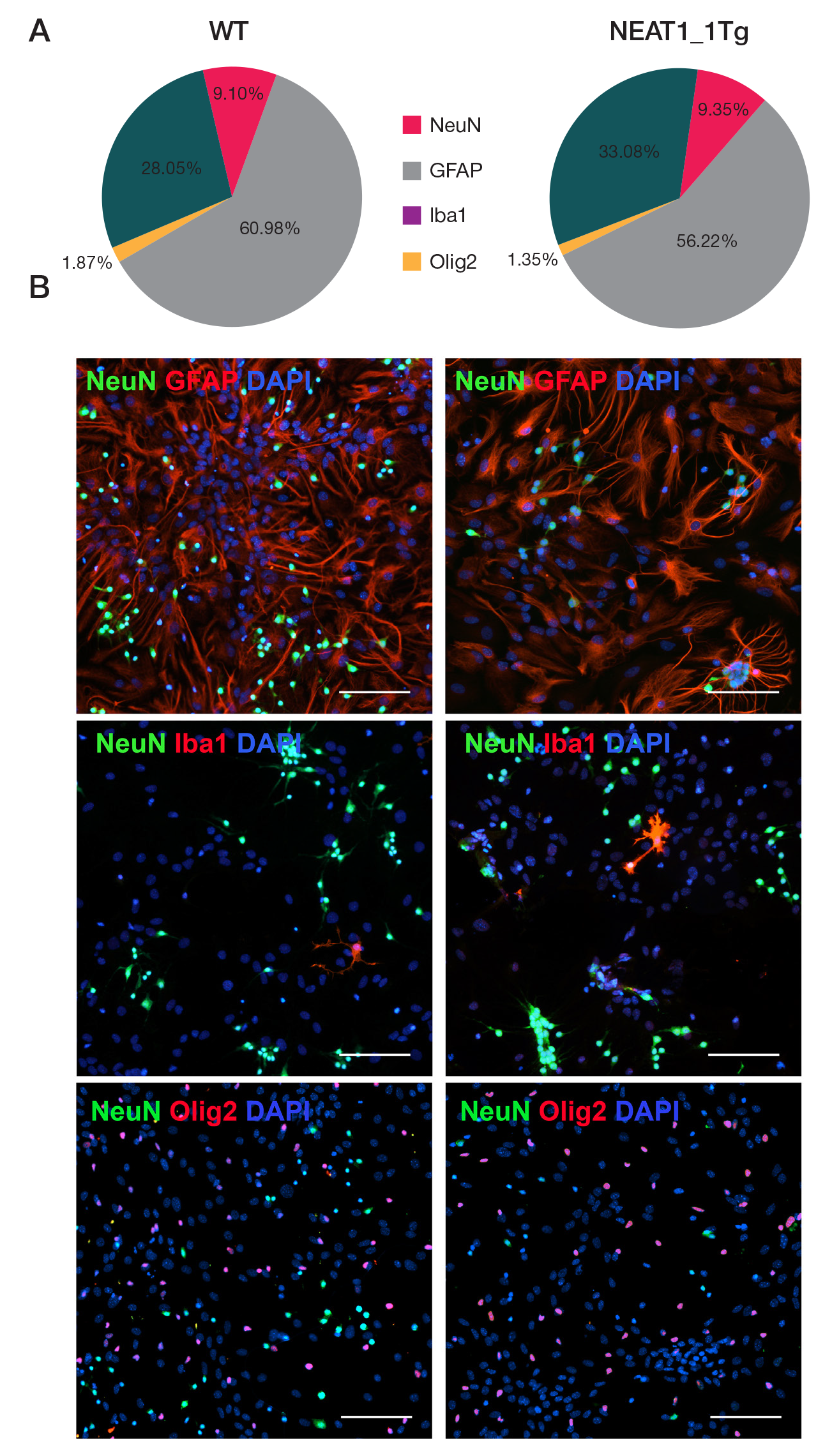
Fig. 1. Cellular composition of hippocampal primary cultures derived from transgenic (NEAT1_1Tg) and wild-type (WT) mice A. Percentages of neurons (NeuN), astrocytes (GFAP), microgliocytes (Iba1), and oligodendrocytes (Olig2) in primary cultures. B. Representative photographs of primary cultures stained for markers of different cell types. Cell nuclei are stained with DAPI. The scale bar is 100 μm
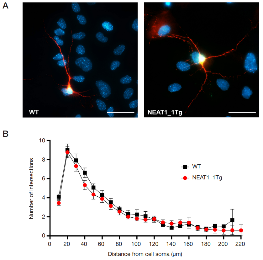
Fig. 2. Morphofunctional characterization of primary neurons. A. Representative photographs of wild-type (WT) and transgenic neurons (NEAT1_1Tg). Immunocytochemical staining for microtubule-associated protein Tau (shown in red), neuronal marker NeuN (shown in green), and cell nuclei stained with DAPI. The scale bar is 50 μm. B. Analysis of branching of primary neuron processes (Sholl-analysis). The graph shows the average number of intersections of circles of a given radius with processes. Multiple t-test with Holm-Sidak correction
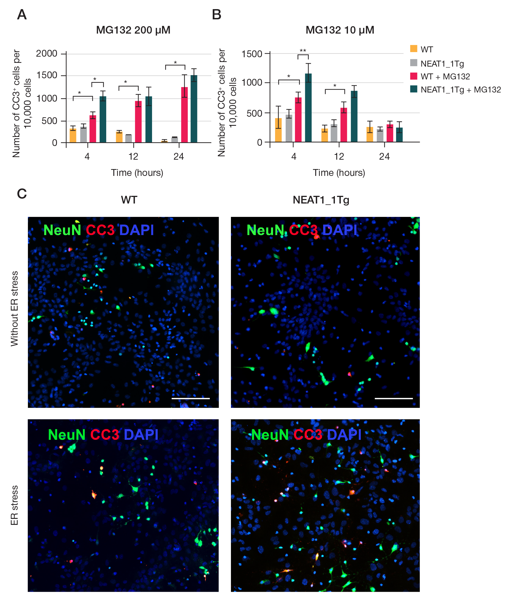
Fig. 3. Apoptotic death in transgenic (NEAT1_1Tg) primary cultures and wild-type (WT) cultures. A. Apoptosis in cultures when treated with 200 μM MG132. B. Apoptosis in cultures when treated with 10 μM MG132. Two-factor analysis of variance, Fisher's test. * — p < 0.05; * * — p < 0.01. C. Representative photographs of cell cultures upon treatment with 10 μM MG132 after 12 h. Neurons stained for the differentiated neuron marker NeuN (shown in green), the apoptotic cell death marker activated caspase 3, CC3 (shown in red), and nuclei stained with DAPI (shown in blue). Scale section is 100 μm
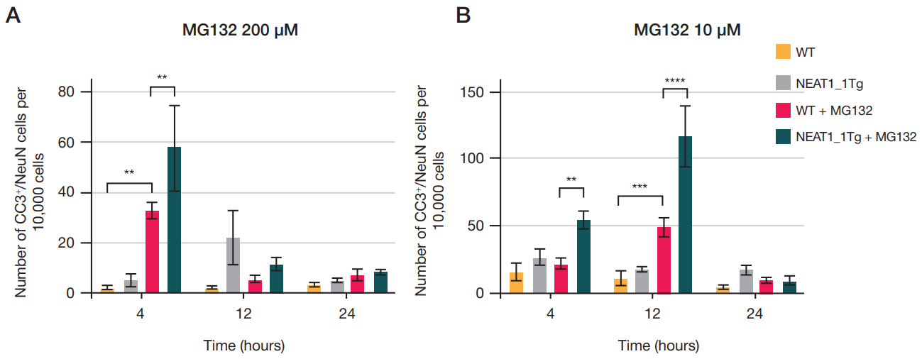
Fig. 4. Apoptotic neuronal death in transgenic (NEAT1_1Tg) primary and wild-type (WT) cultures. A. Apoptosis in cultures treated with 200 μM MG132. B. Apoptosis in cultures treated with 10 μM MG132. Two-way analysis of variance, Fisher's test. * * — p < 0.01; * * * — p < 0.001; * * * * — p < 0.0001
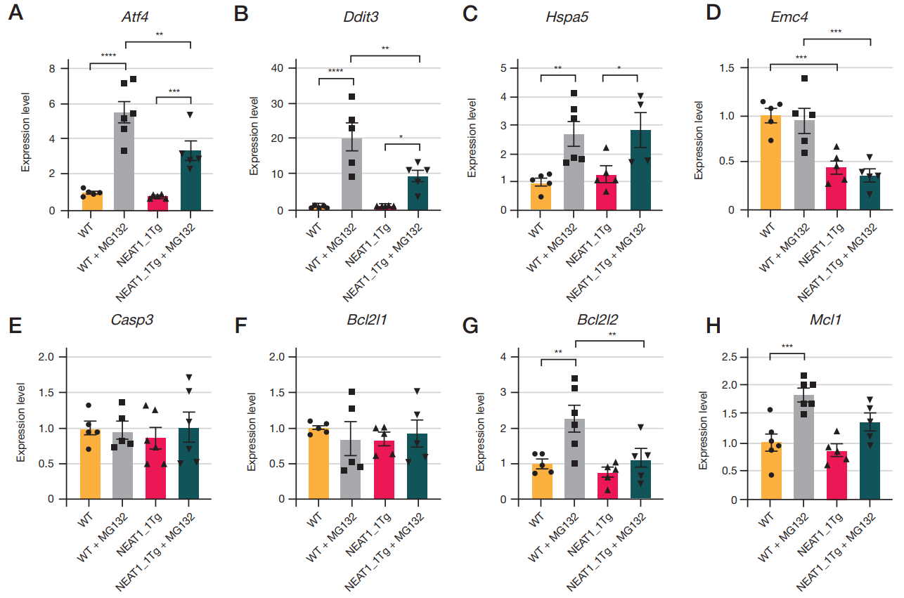
Fig. 5. Gene expression changes in transgenic (NEAT1_1Tg) primary and wild-type (WT) cultures treated with 10 μM MG132. A–G. Expression of genes associated with ER stress. D–Z. Expression of apoptosis-related genes. One-way analysis of variance, Fisher's test. * — p < 0.05; * * — p < 0.01, * * * — p < 0.001; * * * * — p < 0.0001
Table. Sequences of primers used for RT-qPCR