ISSN Print 2500–1094
ISSN Online 2542–1204
BIOMEDICAL JOURNAL OF PIROGOV UNIVERSITY (MOSCOW, RUSSIA)

1 Institute of Gene Biology, Russian Academy of Sciences, Moscow, Russia
2 Joint Institute for High Temperatures of the Russian Academy of Sciences, Moscow, Russia
Correspondence should be addressed: Marina V. Kubekina
Vavilova, 34/5, 119334, Moscow, Russia; moc.liamg@ymukyram
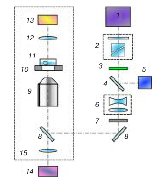
Funding: the procedures involving manipulating embryos using a laser and assessment of expression of the genes responsible for synthesis of heat shock proteins were supported by RSF (project 23-19-00424) and conducted using the equipment of the UNU "Laser Terawatt Femtosecond Complex", which was a part of the Center for Collective Usage "Laser Femtosecond Complex" of the Joint Institute for High Temperatures RAS. The procedures to acquire embryos were supported by the UNU “Transgenbank” grant (№ 075-15-2021-668 of July 29, 2021).
Author contribution: Kubekina MV — immunofluorescence staining and assessment of the heat shock protein expression levels, manuscript writing; Filatov MA — handling embryos, statistical processing, manuscript writing; Silaeva YuYu — general management of the experiment; Sitnikov DS — laser miscrosurgery, data processing, manuscript writing; all authors — discussion and manuscript editing.
Compliance with ethical standards: the study was approved by the Ethics Committee of the Institute of Gene Biology RAS (protocol № 1 dated 25 September 2023) and conducted in strict compliance with the provisions of the Directive 2010/63/EU of the European Parliament and of the Council of 22 September 2010 on the protection of animals used for scientific purposes.