ORIGINAL RESEARCH

Effects of biocompatible piezoelectric membranes on the development of fibrosis associated with the oral mucosal wound regeneration

About authors

1 Siberian State Medical University, Tomsk, Russia

2 Tomsk Polytechnic University, Tomsk, Russia

3 Montana State University, Bozeman, MT, USA

Correspondence should be addressed: Anastasiia D. Koniaeva
Moskovskij trakt, 2, Tomsk, 634034, Russia; moc.liamg@59aynokaysa

About paper

Funding: the study was supported by the RSF (research project No. 23-25-00346)

Author contribution: Koniaeva AD, Varakuta EYu, Bolbasov EN, Stankevich KS — study concept and design; Koniaeva AD, Leiman AE, Kormashov GM, Fedosova MV — data acquisition and processing; Koniaeva AD, Varakuta EYu — manuscript writing; Koniaeva AD, Varakuta EYu, Bolbasov EN, Stankevich KS — manuscript editing.

Compliance with ethical standards: the study was approved by the IACUC of the Siberian State Medical University (protocol No. 11-1 dated 12 July 2022). Rats were handled in accordance with the Directive 2010/63/EU of the European Parliament and of the Council on the protection of animals used for scientific purposes dated 22 September 2010.
Received: 2024-06-18 Accepted: 2024-08-06 Published online: 2024-08-26
|
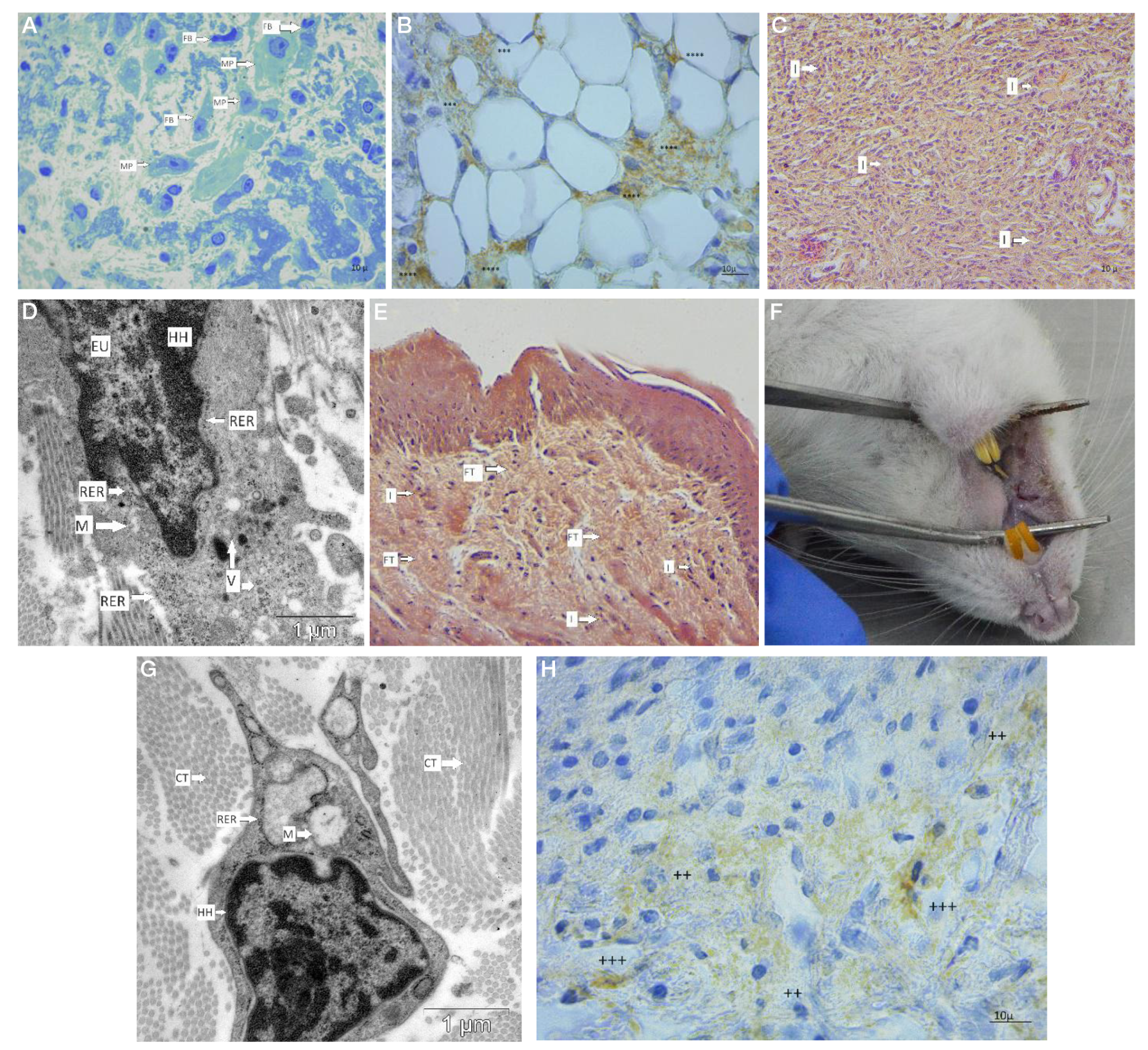
Fig. 1. Phases of the oral mucosal wound defect regeneration. А. Granulation tissue containing macrophages and fibroblasts at the wound defect site (day 3, group 2; ×900; stain: toluidine blue). B. TGFβ1 expression by the granulation tissue cells at the wound defect site, hematoxylin staining of nuclei (day 3, group 1; 900×). C. Loose fibrous connective tissue at the wound defect site showing extensive cell infiltration (day 7, group 1; stain: hematoxylin and eosin; ×400). D. Differentiated fibroblast (day 7, group 2; TEM, 5000×). E. Dense fibrous connective tissue showing cell infiltration (day 12, group 1; stain: hematoxylin and eosin; 400×). F. Macroscopic features after the wound defect regeneration. Soft tissue loss. Scar, 4.5 mm in length. G. Fibrocyte showing no synthetic activity (day 12, group 2; TEM, 5000×). H. CD68 expression by the connective tissue cells, hematoxylin staining of nuclei (day 12, group 2; 400×). FT — dense fibrous connective tissue, I — cell infiltration, EU — euchromatin, HH — heterochromatin, RER — rough endoplasmic reticulum, М — mitochondrion, V — vesicles, MP — macrophage, FB — fibroblast; ++++ — very strong staining; +++ — strong staining; ++ — moderate staining; + — weak staining; ‒ — no detectable staining
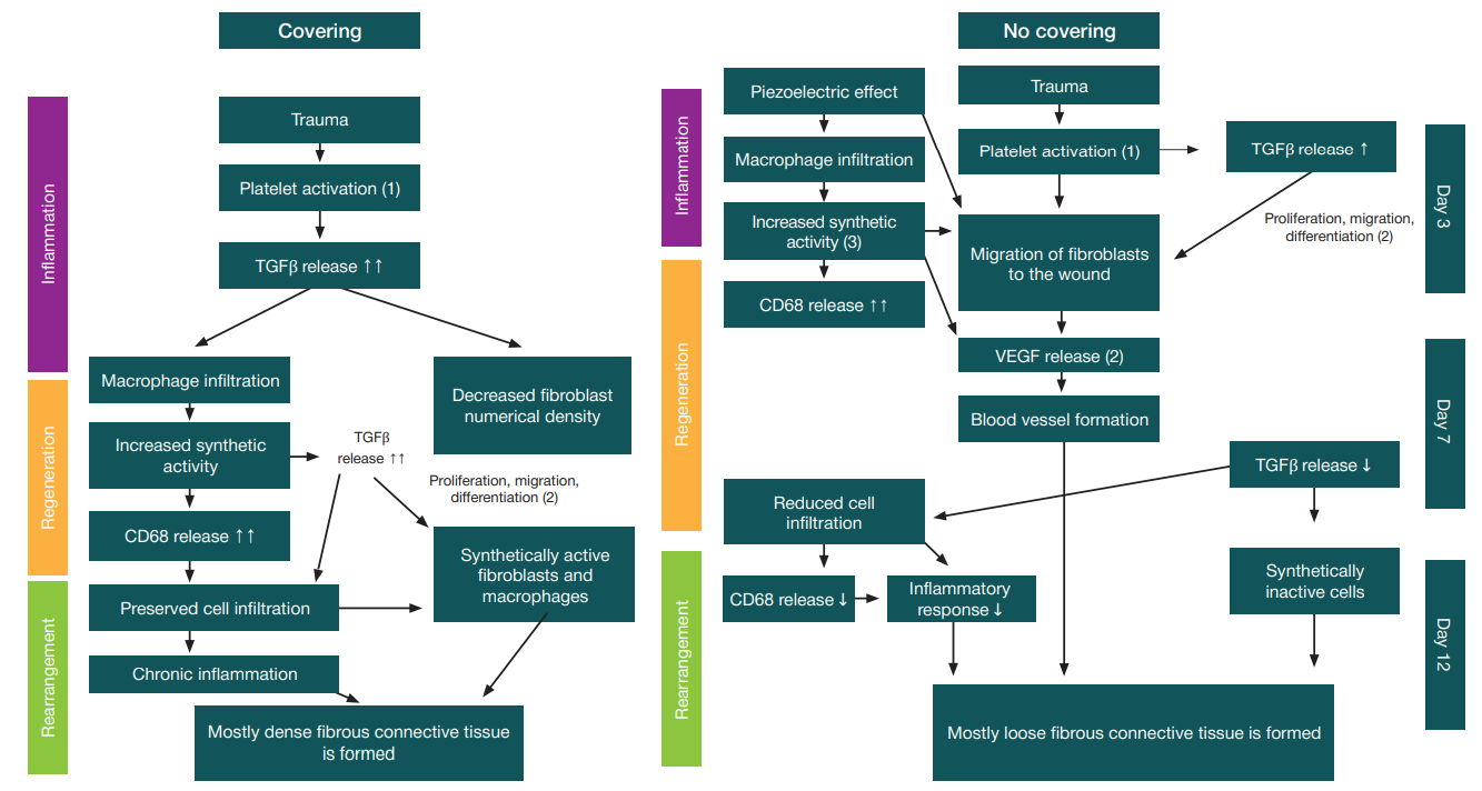
Fig. 2. Scheme of the oral mucosal wound defect regeneration pathomorphogenesis in cases of using/not using a polymeric piezoelectric membrane. ↑ — slightly increased expression; ↑↑ — moderately increased expression; ↑↑↑ — markedly increased expression; ↓ — decreased expression. 1 — according to Morikawa M, Derynck R, et al. [5]; 2 — according to Xu X, Gu SУ, et al. [11]; 3 — according to Zhang T, Wang XF, et al. [12]
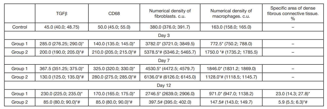
Table. Morphological indicators characterizing the degree of fibrosis in the oral mucosa during regeneration of a wound defect, М(Q1:Q3)
Note: * — significant differences compared to the control group (p < 0.05); # — significant differences compared to group 1 (p < 0.05).