ORIGINAL RESEARCH

Neuroimaging approach to identification of working memory biomarkers in patients with chronic cerebral ischemia

About authors

Research Center of Neurology, Moscow, Russia

Correspondence should be addressed: Vitaly F. Fokin
Volokolamskoye shosse 80, Moscow, 125367, Russia; ur.liam@fvf

About paper

Funding: the study was supported by the RSF grant (No. 22-15-00448).

Author contribution: Fokin VF — manuscript writing; Ponomareva NV — design of physiological and neuropsychological tests, general study design; Konovalov RN — neuroimaging test design; Medvedev RB — Doppler tests; Boravova AI — psychophysiological tests; Lagoda OV — clinical tests; Krotenkova MV — neuroimaging test management; Tanashyan MM — clinical test management, general study design.

Compliance with ethical standards: the study was approved by the Ethics Committee of the Research Center of Neurology (protocol No. 5-6/22 dated 1 June 2022). The informed consent was submitted by all study participants.
Received: 2024-08-29 Accepted: 2024-09-16 Published online: 2024-09-30
|
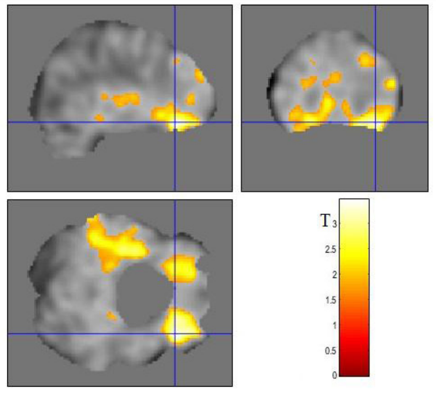
Fig. 1. Brain regions corresponding to the activated voxel difference in patients with high and low working memory indicators based on fMRI data. Lower right — T-value scale. The activated voxel difference at the significance level below 0.05 corresponds to the T-value exceeding 1.68
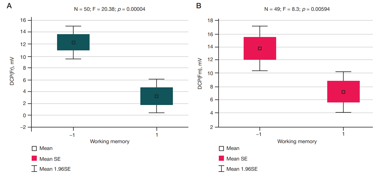
Fig. 2. DCP recorded with the frontal lead in two groups of patients with CCI having low (–1) and high (1) VWM values reported in the resting state (А) and when performing the Luria test (B). Statistical characteristics of differences are shown at the top of the figure. DCP values in mV are along the vertical axis. F — Fisher’s exact test; N — number of subjects; p — significance level
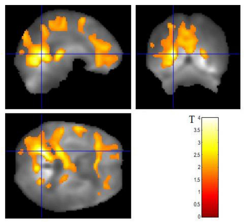
Fig. 3. Brain regions corresponding to the activated voxel difference in CCI patients with low and high DCP in the frontal region. The activated voxels in the right and left lateral fronto-orbital gyri (28 36 –16) and (–28 36 –16), as well as in the left superior temporal gyrus (–40 –17 –7) are highlighted in Fig. 3. Other designations are the same, as in Fig. 1
Fig. 4. Brain regions corresponding to the working memory overlap with the areas that generate infra-slow activity. Interseсtion of the coordinate axes corresponds to the MNI coordinates –28 32 18 — middle frontal gyrus, to the DMN networks and Salience Network. The activated voxel difference in patients with good and worse working memory (А) and with low and high DCPs recorded in the frontal lead of infra-slow potentials (B) is observed in these regions. This area is marked with the coordinate axes intersection on both figures, А and B
Table. Significance of differences in two VWM groups of patients with CCI for various DCP leads