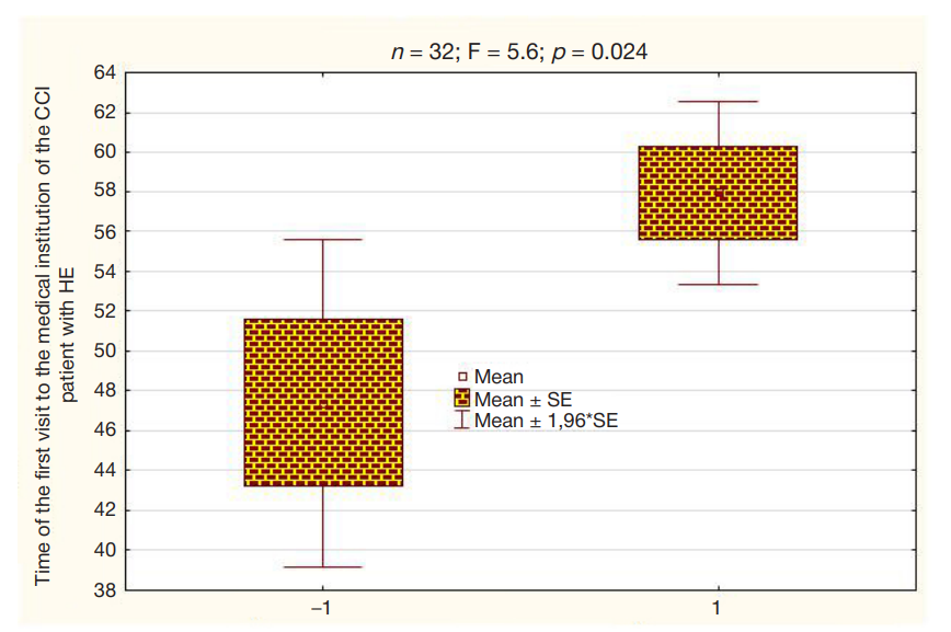
Fig. 1. Value of the cortisol shift associated with cognitive load is related to the time of initial presentation to the medical institution in patients with HE. –1 — average cortisol shift is close to zero (0.02 ± 2.4 nmol/L); 1 — average cortisol shift is 20.09 ± 2.1 nmol/L. On the top there are statistical characteristics of differences in the age of first visit to the medical institution vs. cortisol shift
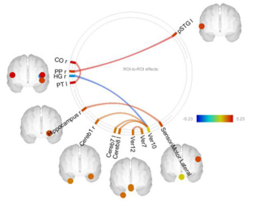
Fig. 2. Connectivity showing significant differences in patients with HE and SE. Red lines are positive, these correspond to higher connectivity values in patients with HE. Blue lines are negative, these correspond to higher connectivity values in patients with SE. Significance was calculated considering multiple comparisons (p-FDR < 0.05). On the right there is a color chart of differences based on the t-test. CO — central operculum cortex, PP — planum polare (part of superior temporal gyrus), HG — Heschl's gyrus, PT — planum temporale, Hippocampus — hippocampus, SensorMotor.Lateral — sensorimotor lateral neural network, STG — superior temporal gyrus, Cereb — cerebellum, Ver — cerebellar vermis, numbers after Cereb and Ver are areas of the cerebellum and vermis, p – posterior, r — right hemisphere, l — left hemisphere
Table 1. Demographic data of women with SE and HE suffering from CCI and significance of differences between the values of these groups
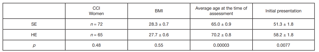
Note: SE — secondary education, HE — higher education, р — significance of differences; the first value is the mean, the second value is the standard error. BMI — body mass index; Average age at the time of assessment — average age of patients at the time of experimental data recording; Initial presentation — average age of the patient’s first referral to the medical institution due to the objectively confirmed CCI symptoms.
Table 2. Effects of education on cortisol levels in women suffering from CCI with higher and secondary education
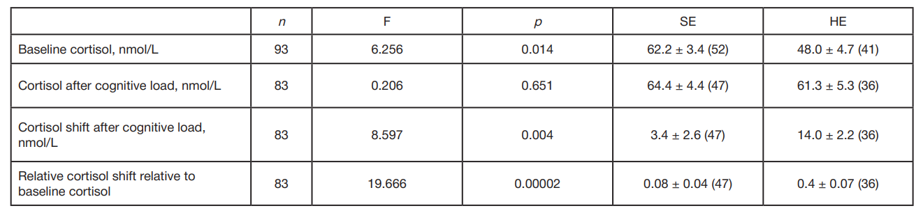
Note: n — number of surveyed individuals, F — Fisher’s exact test, p — significance of differences between the values of two groups; in brackets the number of individuals surveyed; other abbreviations are the same, as in Table 1.
Table 3. Connectivities that are significantly different in the groups of patients with HE and SE and their statistical characteristics
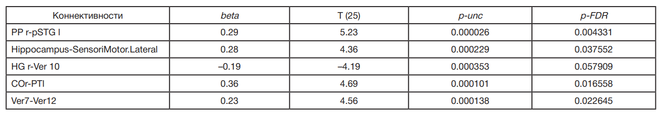
Note: beta — average regression coefficient, Т — T-test, p-unc — uncorrected significance level, p-FDR — significance level considering multiple comparisons. Number of patients — 25 individuals.
Table 4. Correlation coefficients (r) and significance levels (p) for connectivities and cortisol characteristics
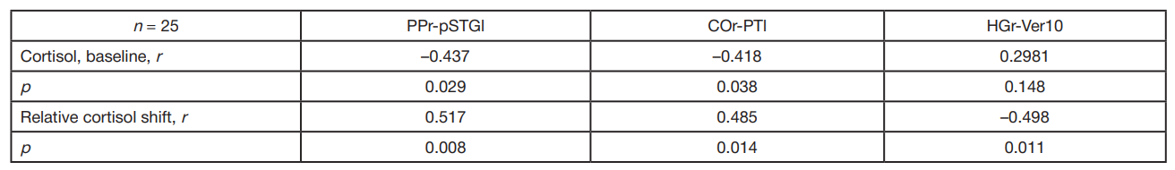
Note: n — number of patients. PP — right planum polare, pSTGl — posterior part of the left superior temporal gyrus; COr — right central operculum cortex, PTl — left planum temporale; HGr — right Heschl’s gyrus, Ver10 — Vermis 10.

