Copyright: © 2025 by the authors. Licensee: Pirogov University.
This article is an open access article distributed under the terms and conditions of the Creative Commons Attribution license (CC BY).

ORIGINAL RESEARCH

Intranasal lipopolysaccharide administration to Sprague-Dawley rats as a biomodel of acute respiratory distress syndrome

Kiseleva VV1, Vishnyakova PA2, Kosyreva AM3, Kananykhina EYu3, Emelianov II1, Elchaninov AV3, Fatkhudinov TH3
About authors

1 Patrice Lumumba Peoples' Friendship University of Russia, Moscow, Russia

2 Kulakov National Medical Research Center for Obstetrics, Gynecology and Perinatology, Moscow, Russia

3 Petrovsky National Research Centre of Surgery, Moscow, Russia

Correspondence should be addressed: Victoria V. Kiseleva
Oparina, 4, Moscow, 117997, Russia; moc.liamg@1991.avosonruk.airotciv

About paper

Funding: the study was supported by the Russian Science Foundation (grant No. 24-25-00203).

Acknowledgements: the authors would like to thank D.A. Areshidze, C. Sci. Biol., Head of the Cellular Pathology Laboratory of the Avtsyn Institute of Human Morphology for performing the complete blood count test in animals.

Author contribution: Kiseleva VV — experimental design and procedure, analysis of the results, manuscript writing; Vishnyakova PA — advice on the experimental procedure, material resources, editing; Kosyreva AM — advice on the experimental procedure, editing; Kananykhina EYu, Emelianov II — animal handling; Elchaninov AV — advice on the experimental procedure, material resources, editing; Fatkhudinov TH — material resources for the study.

Compliance with ethical standards: the study was approved by the Ethics Committee of the Avtsyn Institute of Human Morphology (protocol No. 21 dated 29 March 2019). Animals were handled in accordance to the ARRIVE guidelines and the Directive ЕС 2010/63/EU on the protection of animals used for scientific purposes.
Received: 2025-04-21 Accepted: 2025-05-06 Published online: 2025-05-20
|
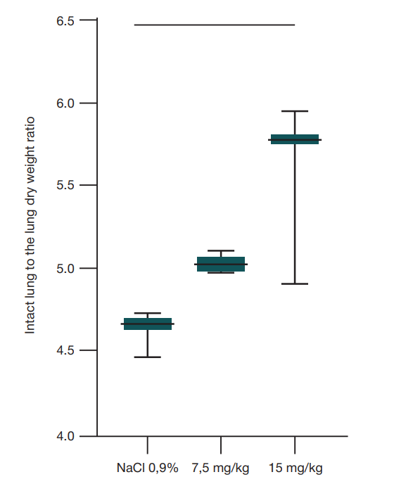
Fig. 1. Results of the lung dry weight determination performed 48 h after the intranasal E. coli O111:B4 LPS administration. Significantly high W/D relative to the control group was determined upon administration of the dose of 15 mg/kg of body weight (p = 0.0228, ordinary one-way Anova)
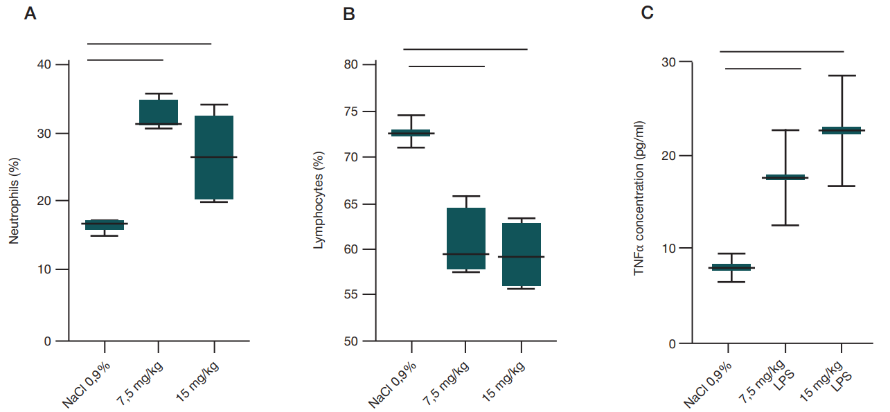
Fig. 2. Complete blood count alterations 48 h after the intranasal LPS administration (А) and serum TNFα concentrations (B). А. Relative lymphocyte counts are decreased compared to the control group (p = 0.0019, ordinary one-way Anova), while neutropil counts are elevated upon administration of both doses (p = 0.0021, ordinary one-way Anova). B. A more than 4-fold increase in serum concentration of the TNFα pro-inflammatory cytokine relative to the control group was observed after the LPS administration, but the differences were non-significant
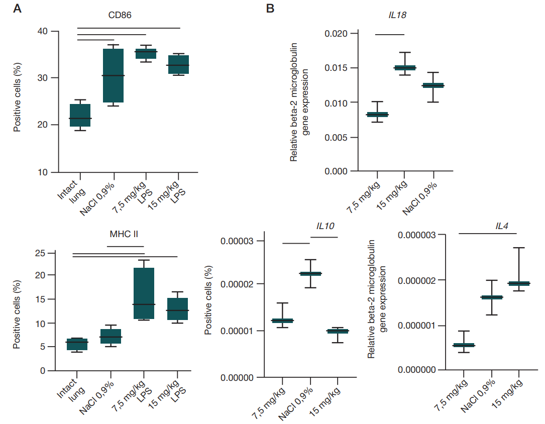
Fig. 3. Lung tissue alterations 48 h after the intranasal LPS administration. А. The counts of cells carrying the marker that is typical for СD86 pro-inflammatory macrophages are elevated relative to the intact lung in all groups (р = 0.0014, ordinary one-way Anova). The counts of cells carrying the marker that is typical for MHC II anti-inflammatory macrophages (p = 0.0050, ordinary one-way Anova) are significantly increased relative to the intact lung after intranasal administration of both LPS doses. The counts of MHC II-positive cells are elevated relative to the group administered saline after LPS administration in a dose of 7.5 mg/kg of body weight. B. When the LPS dose of 15 mg/kg of body weight was administered, there was a significant increase in the IL10 gene expression (ordinary one-way Anova, р = 0.0024) relative to the group administered saline (0.9% NaCl group). IL4 expression decreased (ordinary one-way Anova, р = 0.0194) upon administration of the dose of 7.5 mg/kg of body weight. As for genes IL18 and IL10, the significantly high expression was reported after administration of the LPS dose of 15 mg/kg of body weight (ordinary one-way Anova, р (IL18) = 0.009, р (IL10) = 0.0024)
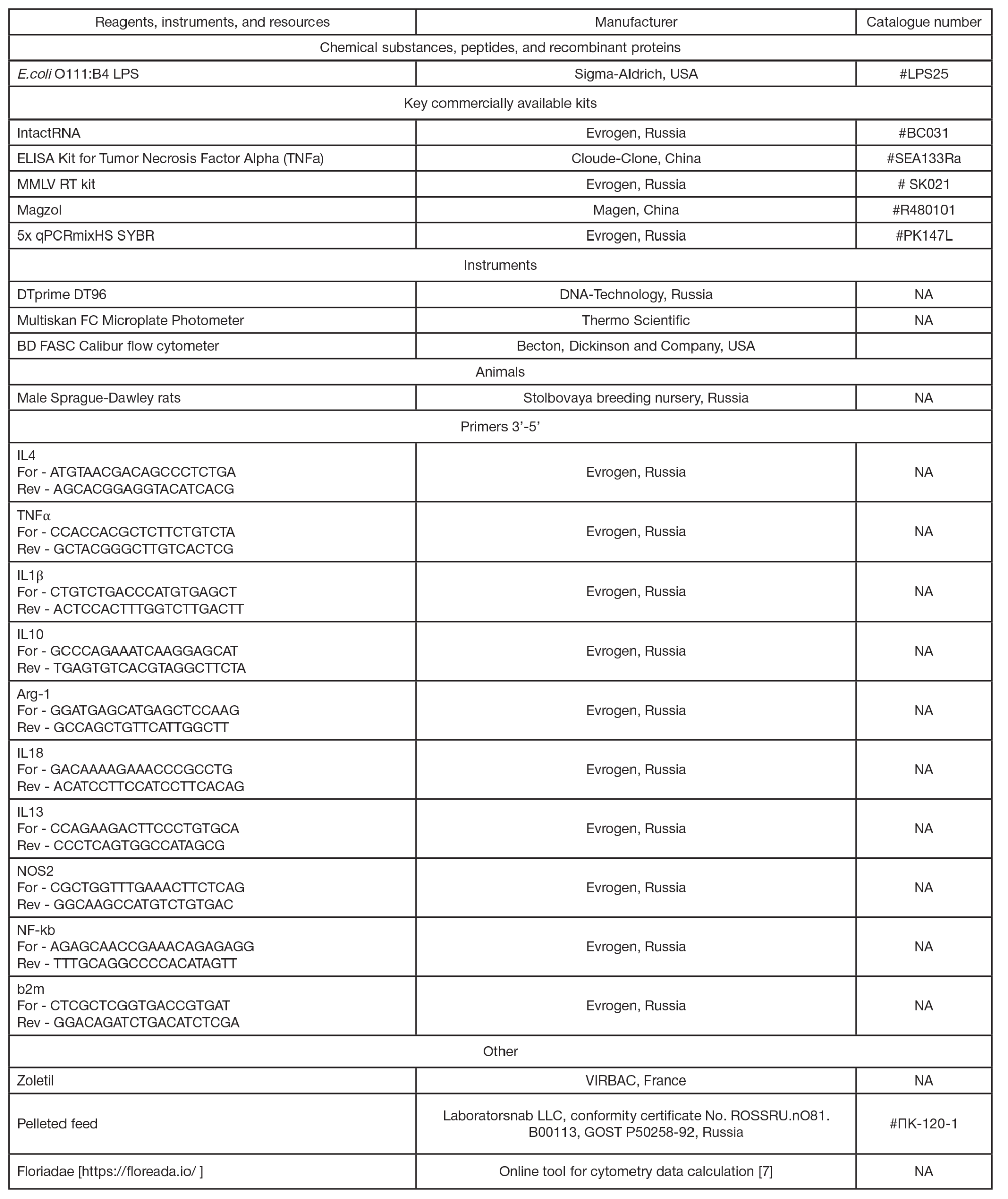
Table 1. Key reagents and materials used in the study
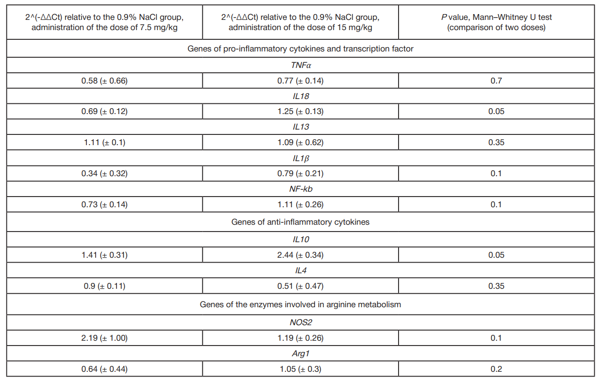
Table 2. Changes in expression of the studied genes in the rat lung after administration of two LPS doses relative to the control group