ISSN Print 2500–1094
ISSN Online 2542–1204
BIOMEDICAL JOURNAL OF PIROGOV UNIVERSITY (MOSCOW, RUSSIA)

1 Research Institute of Translational Medicine, Pirogov Russian National Research Medical University, Moscow, Russia
2 Institute of Biomedical Engineering, MISIS University of Science and Technology, Moscow, Russia
Correspondence should be addressed: Anna V. Ivanova
Ostrovityanova, 1, str.1, Moscow, 117513, Russia; ur.xednay@tirofsof.repus
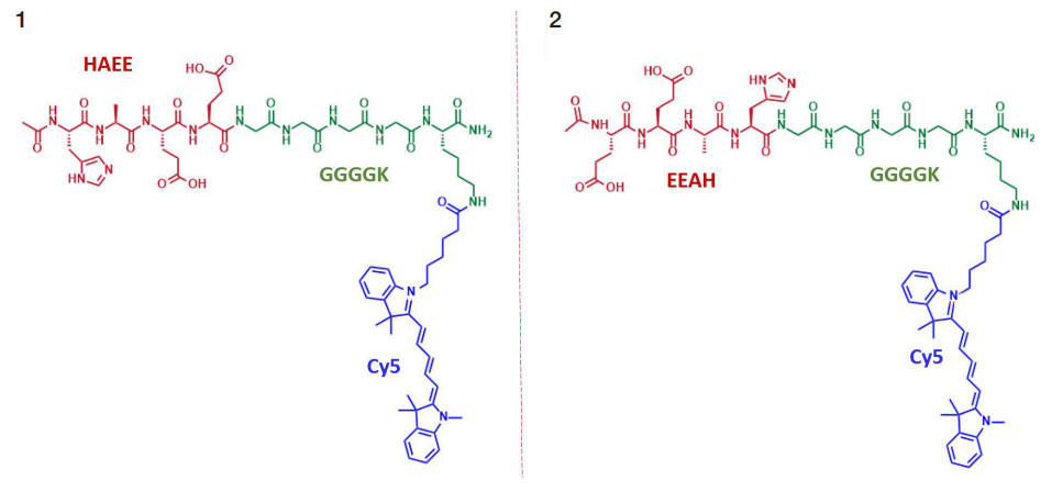
Funding: the work was carried out under the State Assignment "Development of a radiopharmaceutical for the diagnosis of Alzheimer's disease using the HAEE tetrapeptide as a vector molecule", EGISU R&D registration number 1024110600012-8-3.2.25;3.2.26;3.2.12.
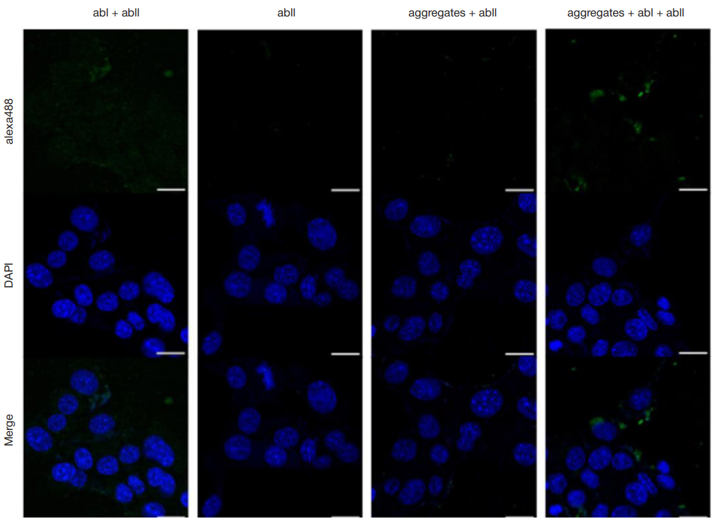
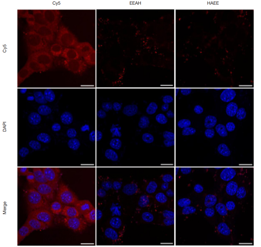
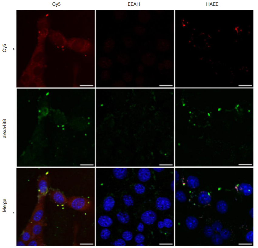
Author contribution: Ivanova AV — literature review, manuscript writing; Chmelyuk NS — fixation, cell sample preparation for microscopic imaging, image acquisition and analysis using a confocal microscope; Kuzmichev IA — synthesis of Ac-HAEEGGGGK(ε-Cy5)-NH2 and Ac-EEAHGGGGK(ε-Cy5)-NH2 fluorescent peptides; Shilyaeva MI — cell sample preparation; Abakumov MA — goal setting, developing the study design, manuscript writing; all the authors contributed to preparation of the paper equally, they confirmed compliance of their authorship with the international ICMJE criteria.