ISSN Print 2500–1094
ISSN Online 2542–1204
BIOMEDICAL JOURNAL OF PIROGOV UNIVERSITY (MOSCOW, RUSSIA)

1 Sechenov First Moscow State Medical University, Moscow, Russia
2 Institute of Theoretical and Experimental Biophysics of the Russian Academy of Sciences, Pushchino, Russia
3 Moscow Polytechnic University, Moscow, Russia
4 Pirogov Russian National Research Medical University, Moscow, Russia
Correspondence should be addressed: Igor L. Kanev
Institutskaya, 3, Pushchino, 142290, Russia; moc.liamg@venak4
Funding: the study was part of a research project sponsored by Neuroconduit LLC and financed under the state assignment No. FFRS-2024-0016 executed by Institute of Theoretical and Experimental Biophysics of the Russian Academy of Sciences.
Acknowledgements: the authors express their deep gratitude for the help of the staff of the Vivarium of Regenerative Medicine of the I. M. Sechenov First Moscow State Medical University, JV Khristidis, BP Ershov, as well as the Laboratory of Digital Microscopic Analysis, AL Fayzulin. Ultrastructure of the materials was examined on equipment of the Research Equipment Sharing Center of Physical Methods for Studying Substances and Materials at the Kurnakov Institute of General and Inorganic Chemistry of the Russian Academy of Sciences.
Author contribution: Gabriyanchik MA — research concept and design, editing; Antonova OY — design development, in vitro testing, data analysis, text preparation; Taylakov ME — production of materials, analysis and statistical data processing, editing; Grachev VA — production of materials, analysis and statistical data processing; Pirogov KS — text preparation; Startseva OI — concept and design of research, editing; Kanev IL — production concept and analysis of the structure of materials, data analysis, text preparation.
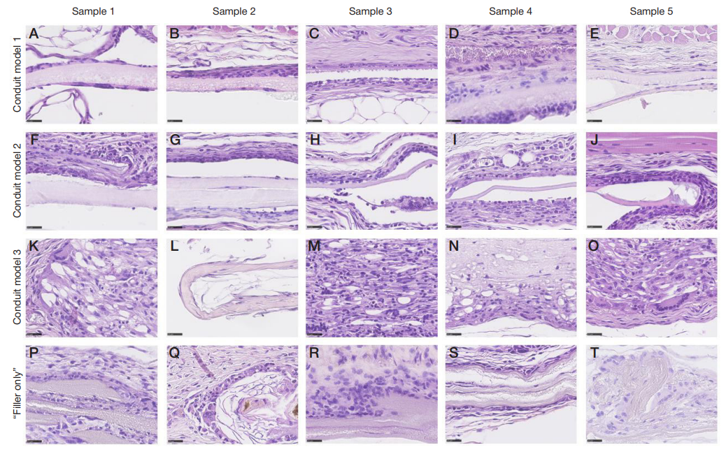
Compliance with ethical standards: the study was approved by the Ethics Committee of the I.M. Sechenov First Moscow State Medical University (Minutes No. 10–25 of April 24, 2025), and conducted in compliance with the provisions of the European Convention for the Protection of Vertebrates used for Experimental and Other Scientific Purposes.