Copyright: © 2026 by the authors. Licensee: Pirogov University.
This article is an open access article distributed under the terms and conditions of the Creative Commons Attribution license (CC BY).

ORIGINAL RESEARCH

Antitumor activity of prototype drugs based on the Newcastle disease virus in the AKATOL tumor model

Yurchenko KS1 , Adamenko LS1 , Kovalenko VS1 , Budko AI2 , Gagloeva TD2 , Timaeva OI2 , Trashkov AP2 , Shestopalov AM1
About authors

1 Federal Research Center of Fundamental and Translational Medicine (FRC FTM), Novosibirsk, Russia

2 Kurchatov Institute, Moscow, Russia

Correspondence should be addressed: Ksenia S. Yurchenko
Timakova, 2, Novosibirsk, 630060, Russia; ur.mtfcrf@oknehcruisk

About paper

Funding: the work was supported under the State Assignment No. 075-00486-26-00 of the Ministry of Science and Higher Education of the Russian Federation.

Author contribution: Yurchenko KS — study concept and design, article authoring, visualization; Adamenko LS — preparation of virus-containing drugs; Kovalenko VS — review and analysis of literature, article authoring; Budko AI — experiments on mice, statistical analysis; Gagloeva TD — experiments on mice, preparation of samples of drugs; Timaeva OI — study concept, text editing; Trashkov AP — study design, experiments on mice, formal analysis, text editing; Shestopalov AM — formulation of study goals and objectives, text editing, project management.

Compliance with ethical standards: the study was approved by the Ethics Committee of the Federal Research Center of Fundamental and Translational Medicine (Minutes No. 26 of September 29, 2026). The animals were kept, cared for, manipulated and removed from the experiment in accordance with the principles of humane treatment of laboratory animals set out in the Directive 86/609/EEC and the Declaration of Helsinki.
Received: 2026-08-27 Accepted: 2026-10-07 Published online: 2026-10-18
|
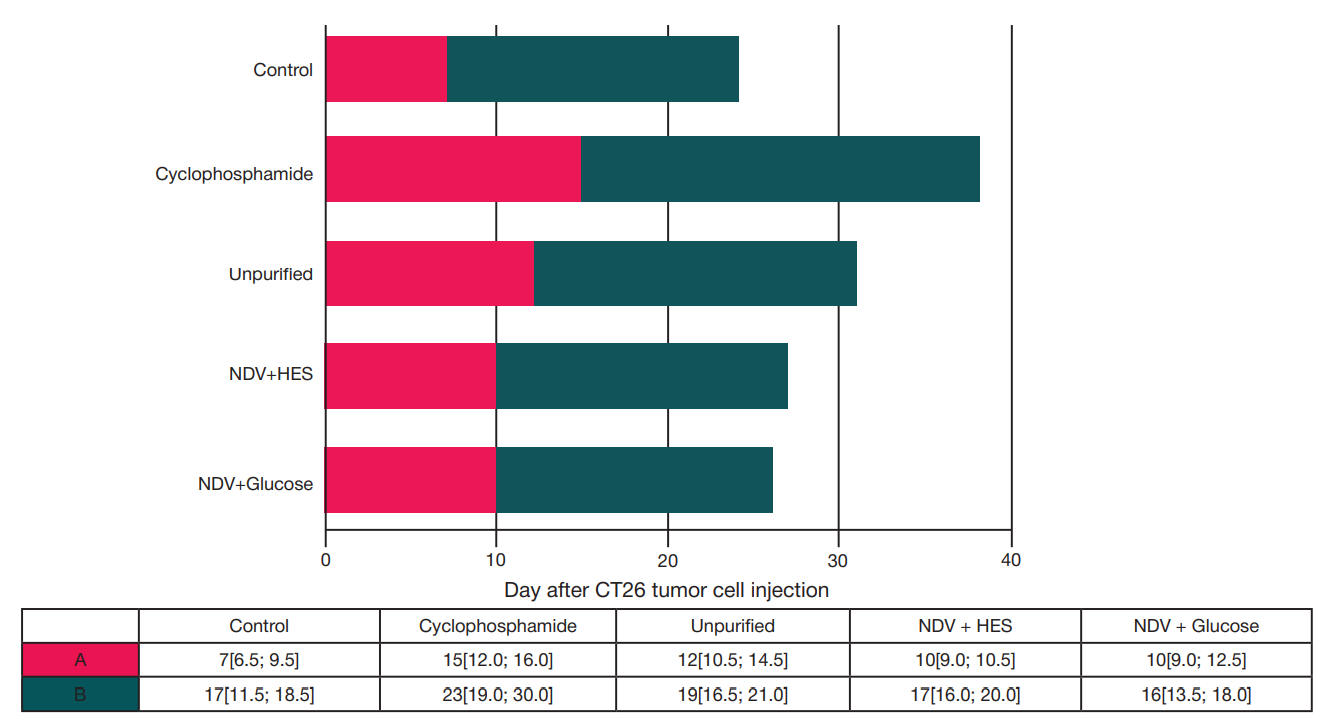
Fig. 1. The time of registration of the primary tumor nodule of subcutaneously transplanted CT26 colon adenocarcinoma after intravenous antitumor therapy (diagram, A), and the duration of the growth period of the tumor nodule from the moment of detection to the death of the animal (diagram, B) (day)
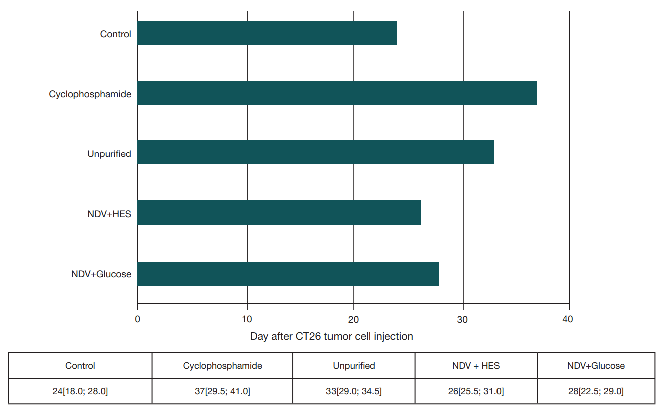
Fig. 2. Mean survival time of BALB/c mice with CT26 colon adenocarcinoma after intravenous antitumor therapy
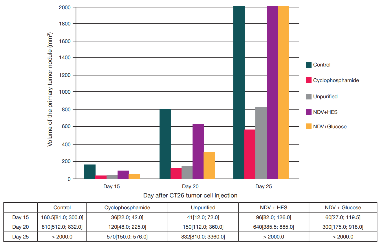
Fig. 3. Dynamics of growth of the primary tumor nodule of CT26 colon adenocarcinoma after intravenous antitumor therapy in the early period after tumor grafting, day after grafting
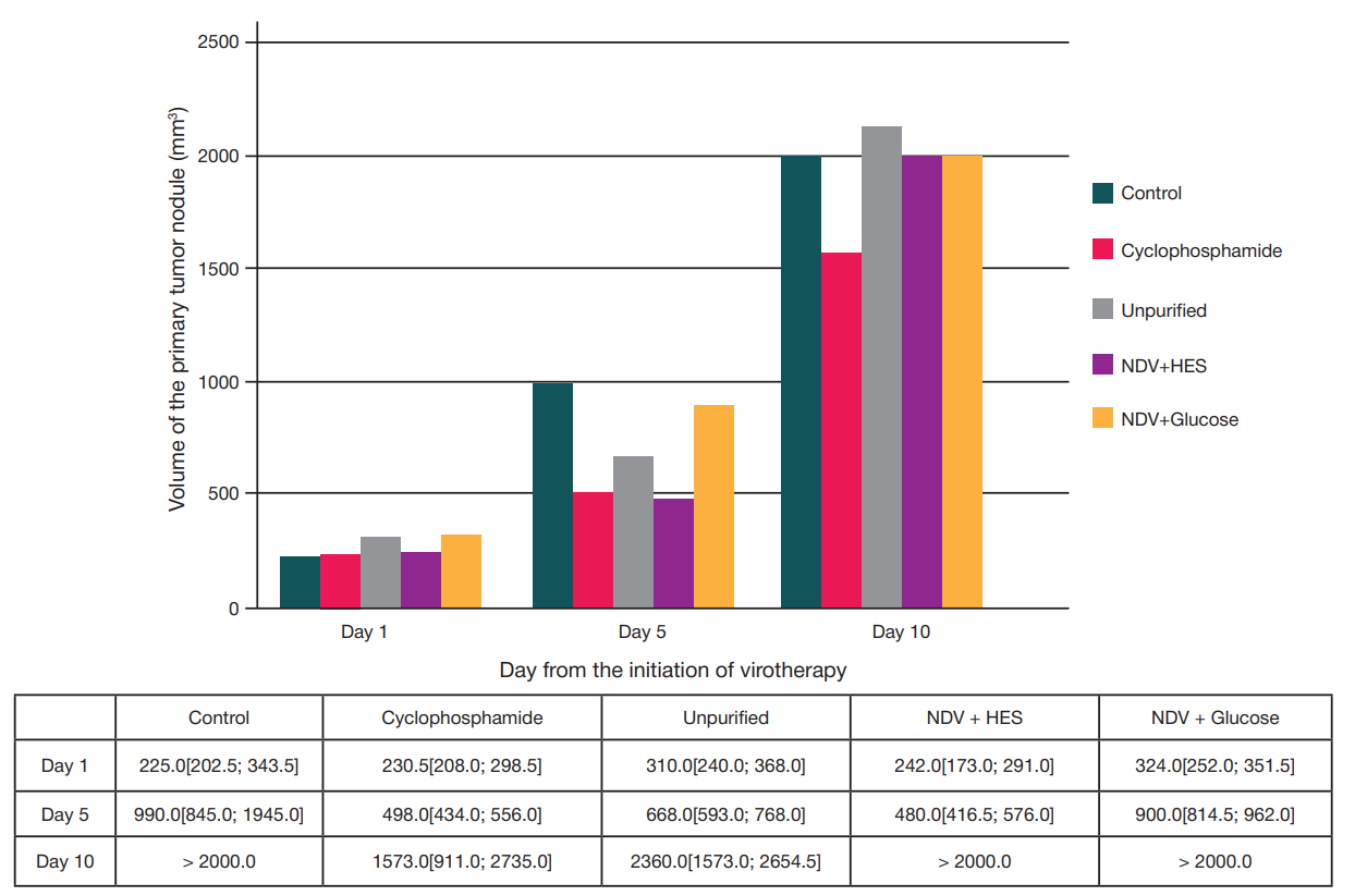
Fig. 4. Dynamics of growth of the primary tumor nodule of CT26 colon adenocarcinoma after antitumor therapy of the established tumor, day after start of therapy
Table. Infectious titers of virus-containing preparations, strain NDV/Altai/pigeon/770/2011
Note: culture-derived — propagated on Vero E6 cell culture; * — aliquots of virus-containing samples for infectious titer analysis were obtained immediately after filtration, ultrafiltration, and addition of HES and glucose solutions, without freezing and long-term storage.