METHOD

Modification of the method for analysis of genome editing results using CRISPR/Cas9 system on preimplantation mouse embryos

About authors

1 Marlin Biotech, Moscow, Russia

2 Institute of Gene Biology, Russian Academy of Sciences, Moscow, Russia

3 Department of Neurology, Neurosurgery and Medical Genetics, Medical Faculty,
Pirogov Russian National Research Medical University, Moscow, Russia

Correspondence should be addressed: Tatiana Dimitrieva
ul. Vavilova, d. 34/5, Moscow, Russia, 119334; moc.liamg@at.aveirtimid

About paper

Acknowledgements: authors thank the Shared Resource Center of the Institute of Gene Biology of Russian Academy of Sciences for the equipment provided for this research.
Received: 2016-06-16 Accepted: 2016-06-21 Published online: 2017-01-05
|
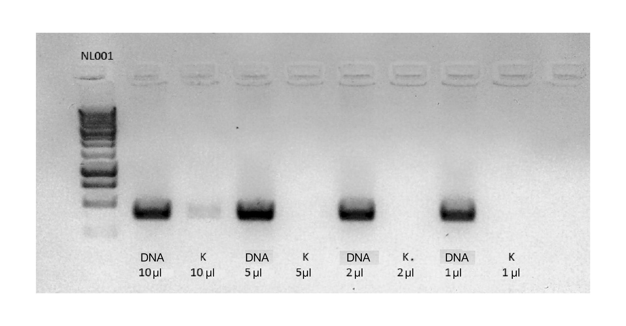
Fig. 1. Amplification of DMD intron 8 using different amounts of matrix
Blastocysts were lysed by Sakurai et al. method [19] with modifications in a final volume of 20 μl. 1 to 10 μl of total DNA solution was used for polymerase chain reaction (PCR). The appropriate volume of water from the final wash, treated similarly was used as the control for each sample. PCR products were separated in a 2 % agarose gel using intercalating dye. NL001 (Evrogen, Russia) was used as the DNA fragment length marker.
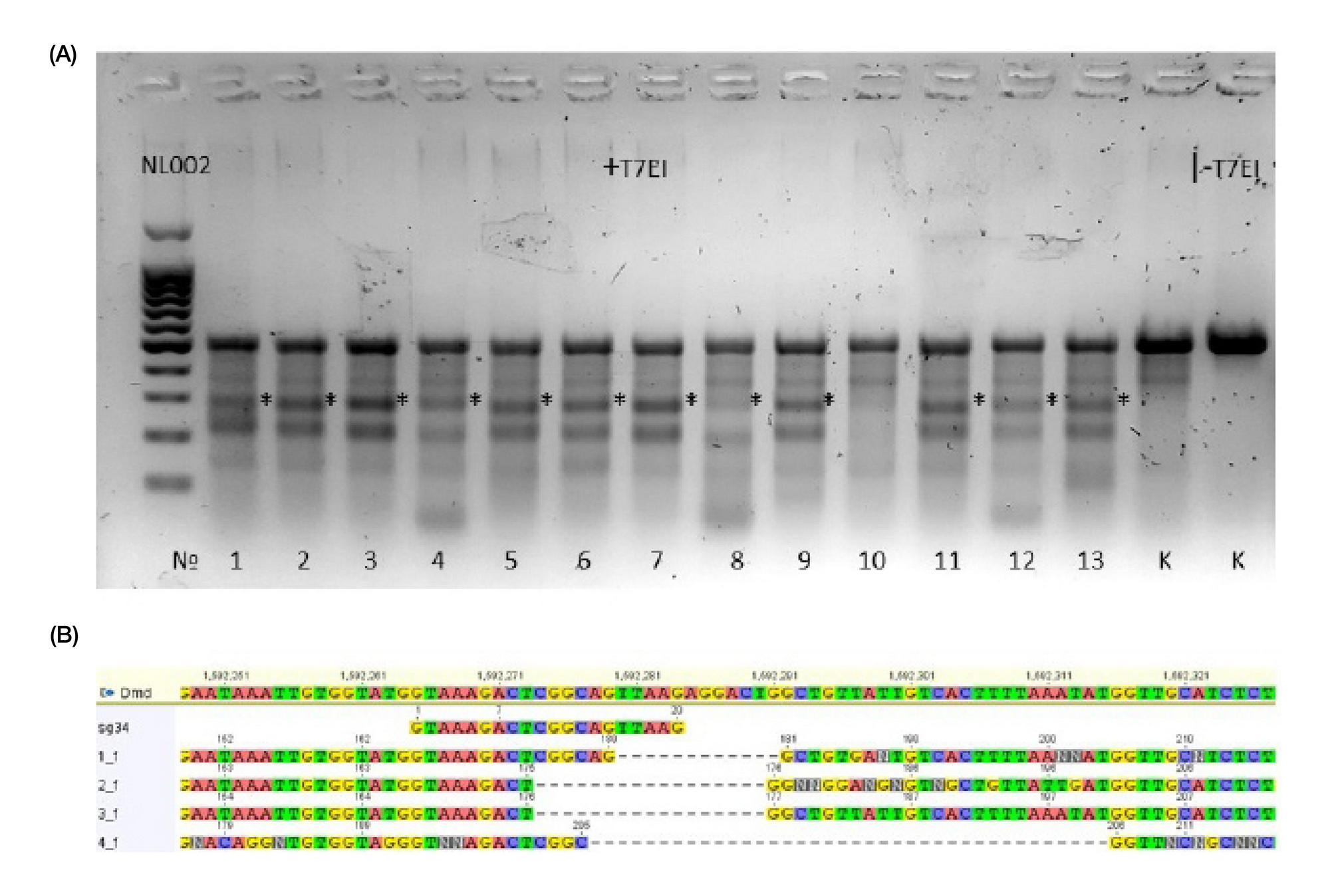
Fig. 2. Detection of insertions and deletions in DMD intron 34 after microinjection of the sg34/Cas9 complex
The intron fragment around recognition site g34 was amplified with injected and control embryos. PCR products were annealed in control amplicon and processed with bacteriophage T7 endonuclease I. Cleavage into two fragments occurred in samples that contained insertions and deletions. Such samples are marked in the figure with an asterisk * (A). The boundaries of insertions and deletions in selected samples were determined via Sanger sequencing (B). PCR products were separated in a 2 % agarose gel using intercalating dye. NL001 (Evrogen, Russia) was used as the DNA fragment length marker.
Fig. 3. Detection of insertion at the break site in DMD intron 8 after microinjection of the sg31/Cas9 complex with repair matrix
The intron fragment around recognition site g34 was amplified with injected and control embryos. PCR products were treated with restriction endonuclease BamHI, whose recognition site was encoded in repair matrix. Cleavage into two fragments occurred in samples that contained insertions and deletions. Such samples are marked in the figure with an asterisk * (А). The presence of an insertion in selected samples was confirmed by Sanger sequencing (B). PCR products were separated in a 2 % agarose gel using intercalating dye. NL001 (Evrogen, Russia) was used as the DNA fragment length marker.
Oligonucleotides sequences