Copyright: © 2018 by the authors. Licensee: Pirogov University.
This article is an open access article distributed under the terms and conditions of the Creative Commons Attribution license (CC BY).

ORIGINAL RESEARCH

The use of antimicrobial photodynamic therapy mediated by MC540 in the infected wound model

Shmigol TA1, Sobianin KA2, Prusak-Glotov MV1, Shchelykalina SP3, Nevezhin EV3, Yermolaeva SA4, Negrebetsky VV1
About authors

1 Department of Medicinal Chemistry and Toxicology,
Pirogov Russian National Research Medical University, Moscow

2 Laboratory of Biological Research. Institute for Translational Medicine,
Pirogov Russian National Research Medical University, Moscow

3 Department of Medical Cybernetics and Informatics, Biomedical Faculty,
Pirogov Russian National Research Medical University, Moscow

4 Laboratory for the Ecology of Pathogens,
Gamaleya Federal Research Center for Epidemiology and Microbiology, Moscow

Correspondence should be addressed: Tatiana Shmigol
Ostrovityanova 1, Moscow, 117997; moc.liamg@hsithsitat

About paper

Funding: this study was supported by the Russian Foundation for Basic Research (Project ID 16-33-00970 mol_a).
Received: 2018-01-20 Accepted: 2018-03-23 Published online: 2018-04-28
|
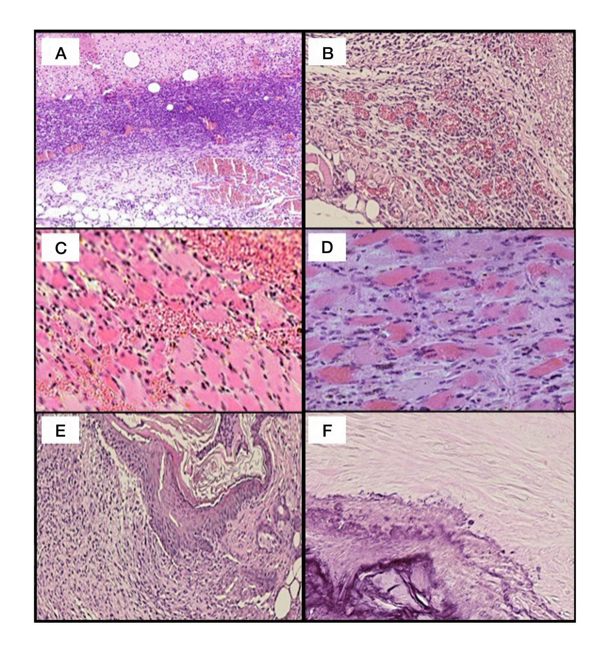
Fig. 1. Microscopy of infected wounds of mice after PDT. Stain used: hematoxylin and eosin (А) Immature granulation tissue on day 4, ×150. (B) Vascular hyperemia and fluid accumulation around the capillary bed on day 4, ×350. (C) Blood in the muscle tissue on day 4, ×250. (D) Fragments of muscle tissue showing intracellular cell regeneration of day 7, ×450. (E) Epithelialization and scarring on day 14, ×150. (F) Hyalinized and calcified scar tissue on day 36, ×150
Table 1. Study design
Table 2. Bacterial contamination of the wound measured on different days of the experiment, CFU/ml
Table 3. Morphometry of wound matrix remodeling in BALB/c mice
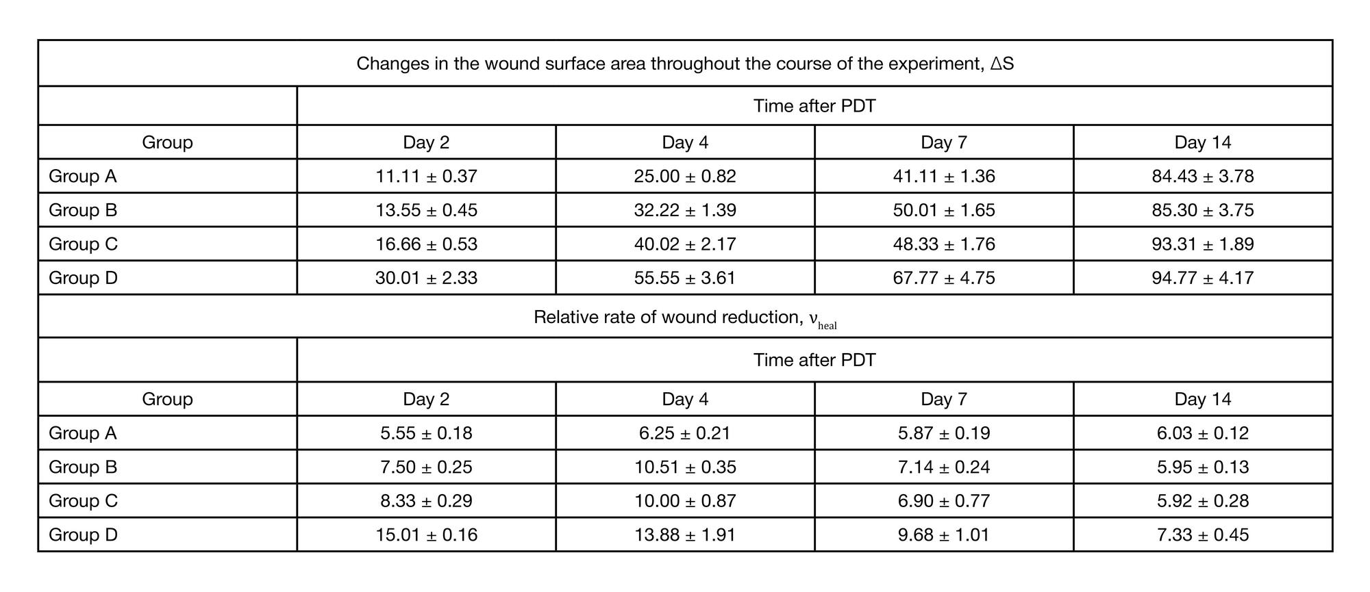
Table 4. Rates of epithelial repair of infected wounds in BALB/c mice